. 2016 Aug 25;13(1):197.
doi: 10.1186/s12974-016-0663-y.

Cortisol-induced immune suppression by a blockade of lymphocyte egress in traumatic brain injury

Affiliations

Cortisol-induced immune suppression by a blockade of lymphocyte egress in traumatic brain injury

Tingting Dong et al. J Neuroinflammation. .

Abstract

Background: Acute traumatic brain injury (TBI) represents one of major causes of mortality and disability in the USA. Neuroinflammation has been regarded both beneficial and detrimental, probably in a time-dependent fashion.

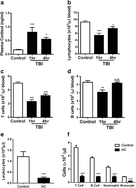
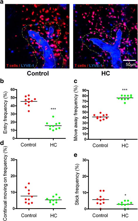
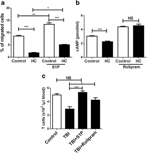
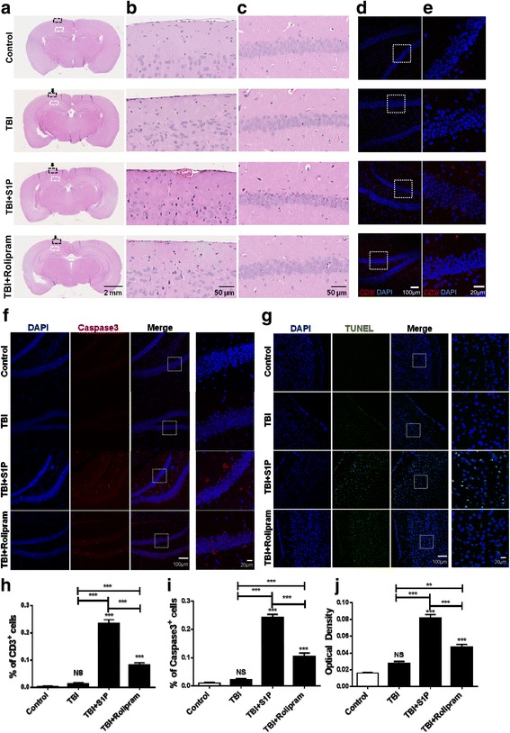
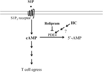
Methods: To address a role for neuroinflammation in brain injury, C57BL/6 mice were subjected to a closed head mild TBI (mTBI) by a standard controlled cortical impact, along with or without treatment of sphingosine 1-phosphate (S1P) or rolipram, after which the brain tissue of the impact site was evaluated for cell morphology via histology, inflammation by qRT-PCR and T cell staining, and cell death with Caspase-3 and TUNEL staining. Circulating lymphocytes were quantified by flow cytometry, and plasma hydrocortisone was analyzed by LC-MS/MS. To investigate the mechanism whereby cortisol lowered the number of peripheral T cells, T cell egress was tracked in lymph nodes by intravital confocal microscopy after hydrocortisone administration.

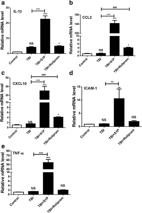
Results: We detected a decreased number of circulating lymphocytes, in particular, T cells soon after mTBI, which was inversely correlated with a transient and robust increase of plasma cortisol. The transient lymphocytopenia might be caused by cortisol in part via a blockade of lymphocyte egress as demonstrated by the ability of cortisol to inhibit T cell egress from the secondary lymphoid tissues. Moreover, exogenous hydrocortisone severely suppressed periphery lymphocytes in uninjured mice, whereas administering an egress-promoting agent S1P normalized circulating T cells in mTBI mice and increased T cells in the injured brain. Likewise, rolipram, a cAMP phosphodiesterase inhibitor, was also able to elevate cAMP levels in T cells in the presence of hydrocortisone in vitro and abrogate the action of cortisol in mTBI mice. The investigation demonstrated that the number of circulating T cells in the early phase of TBI was positively correlated with T cell infiltration and inflammatory responses as well as cell death at the cerebral cortex and hippocampus beneath the impact site.

Conclusions: Decreases in intracellular cAMP might be part of the mechanism behind cortisol-mediated blockade of T cell egress. The study argues strongly for a protective role of cortisol-induced immune suppression in the early stage of TBI.

Keywords: Cortisol; Inflammation; T lymphocytes; TBI; cAMP.


Figures

Fig. 1
Fig. 1
Inverse relationship between cortisol and lymphocytes in blood following TBI. a Plasma cortisol was quantified before and 1 and 4 h after TBI. In parallel, the numbers of peripheral lymphocytes (b), T cells (c), and B cells (d) were analyzed at the same time points. A total number of leukocytes (e) or indicated cells (f) were measured in blood by flow cytometry 4 h after i.p. injection of 10 mg/kg hydrocortisone. Data are expressed as means ± SEM. n = 5 in (a) or 6 in (b, c, d, e, f). Significance was determined using one-way ANOVA (a, b, c, d) or non-parametric Mann-Whitney t test (e, f). *P < 0.05, **P < 0.01, ***P < 0.001, and NS, no significance compared before and after TBI or HC treatment. The experiment was repeated three times with similar results
Fig. 2
Fig. 2
T cell egress is blocked by hydrocortisone. The representative images taken from control or hydrocortisone (HC)-treated mice are shown in (a). LYVE-1+ cortical sinuses are shown in blue pseudocolor in order to distinguish them with CMTMR labeled T cells (red) and the representative sinus area is delineated by a dotted white line. The dotted yellow line outlines the area within 30 μm of distance from the outer boundaries of cortical sinuses. Note: few T cells within cortical sinus in the presence of HC. Scale bar, 50 μm. Frequencies at which T cells entered (b), moved away (c), crawled on (d), or stuck to (e) (kept adhering to one point on the sinus wall and never displaced during the imaging period after they engaged the sinus) the cortical sinuses in control and HC-treated mice were calculated by manually tracking individual cells in each time-lapse image, with a total of 200 cells randomly selected in 10~15 imaging stacks. Each dot represents data from a single time-lapse image, and bars represent the means. Significance was measured using non-parametric Mann-Whitney t test. *P < 0.05, ***P < 0.001 in the presence or absence of hydrocortisone. Data are combined from two independent experiments each with two lymph nodes imaged in each treatment. The experiment was repeated two times with similar results
Fig. 3
Fig. 3
S1P or rolipram increases peripheral T cells in TBI mice. a T cell migration was analyzed in 48-well micro chemotaxis chamber, with 20 μM hydrocortisone or vehicle in the upper chamber and 20 nM S1P or vehicle in the lower chamber. The number of migrated cells was assessed 4 h later in the lower chambers. b T cells were pretreated with 10 μM rolipram or saline for 15 min and then with 100 μM hydrocortisone or vehicle treatment for 5 min, after which intracellular cAMP level was measured. c Peripheral T cells were measured before and 1 h after TBI. S1P or rolipram was i.p. injected immediately after TBI. Results are expressed as means ± SEM. n = 9 for (a), 6 for (c), or 4 for (b). Significance was determined using two- (a, b) or one-way (c) ANOVA. *P < 0.05, **P < 0.01, ***P < 0.001, and NS, no significance compared between indicated groups. The experiment was repeated three times with similar results
Fig. 4
Fig. 4
S1P or rolipram exaggerates inflammatory responses in injured brain. IL-1β (a), CCL2 (b), CXCL10 (c), ICAM-1 (d), and TNF-α (e) were analyzed at the impact site of the cerebral cortex in 3 days after TBI by qRT-PCR. The data are expressed as means ± SEM and normalized to β-actin. n = 5, significance was measured using one-way ANOVA. *P < 0.05, **P < 0.01, ***P < 0.001 and NS, no significance compared between indicated groups. ### P < 0.001 compared between TBI and TBI + rolipram in CCL2 and CXCL10 expression level by non-parametric Mann-Whitney t test. The experiment was repeated three times with similar results
Fig. 5
Fig. 5
A protective role for cortisol in TBI pathogenesis. a Histologic examination of normal control and injured brain at 7 days after TBI with or without administration of S1P or rolipram. The impact site was pointed by an arrow. The region of the cerebral cortex was highlighted in a dashed black line square and enlarged in panel (b); and the hippocampus was outlined by a dashed white line square and magnified in panel (c). Representative results of six mice in each group. d Representative immunofluorescence results of anti-CD3 antibody staining at hippocampus beneath the injured site and enlarged in panel (e). f Representative immunofluorescence staining for Caspase-3 expression at hippocampus beneath the injured site. g Representative TUNEL staining for apoptosis cells at the injury site. Percentages of CD3-positive cells in panel (e), Caspase-3-positive cells in panel (f), and optical density of TUNEL staining in panel (g) were determined by ImageJ and expressed as means ± SEM in (h), (i), or (j), respectively. n = 6, significance was measured using one-way ANOVA. *P < 0.05, **P < 0.01, ***P < 0.001 and NS, no significance compared between indicated groups. The experiment was repeated three times with similar results
Fig. 6
Fig. 6
Schematic illustration of a possible mechanism underlying cortisol-mediated blockade of T cell egress. cAMP is one of the important second messengers downstream the S1P1 receptor and its production takes central part in the control of T cell egress. One of the cortisol (HC) activities may activate cAMP phosphodiesterase (PDE4) either directly or indirectly and enhance degradation of cAMP to 5′-AMP. Cortisol-facilitated degradation of cAMP may be one of the mechanisms where cortisol compromises T cell egress in the presence of S1P. On the contrary, rolipram inhibits PDE4, leading to increased levels of cAMP and promoting T cell egress

References

    1. Selassie AW, Zaloshnja E, Langlois JA, Miller T, Jones P, Steiner C. Incidence of long-term disability following traumatic brain injury hospitalization, United States, 2003. J Head Trauma Rehabil. 2008;23(2):123–31. doi: 10.1097/01.HTR.0000314531.30401.39. - DOI - PubMed
    1. Bramlett HM, Dietrich WD. Progressive damage after brain and spinal cord injury: pathomechanisms and treatment strategies. Prog Brain Res. 2007;161:125–41. doi: 10.1016/S0079-6123(06)61009-1. - DOI - PubMed
    1. Marklund N, Bakshi A, Castelbuono DJ, Conte V, McIntosh TK. Evaluation of pharmacological treatment strategies in traumatic brain injury. Curr Pharm Des. 2006;12(13):1645–80. doi: 10.2174/138161206776843340. - DOI - PubMed
    1. Finnie JW. Neuroinflammation: beneficial and detrimental effects after traumatic brain injury. Inflammopharmacology. 2013;21(4):309–20. doi: 10.1007/s10787-012-0164-2. - DOI - PubMed
    1. Ziv Y, Ron N, Butovsky O, Landa G, Sudai E, Greenberg N, et al. Immune cells contribute to the maintenance of neurogenesis and spatial learning abilities in adulthood. Nat Neurosci. 2006;9(2):268–75. doi: 10.1038/nn1629. - DOI - PubMed

Publication types

MeSH terms