

Latent mitochondrial DNA deletion mutations drive muscle fiber loss at old age
- PMID: 27561813
- PMCID: PMC5114866
- DOI: 10.1111/acel.12520
Latent mitochondrial DNA deletion mutations drive muscle fiber loss at old age
Abstract
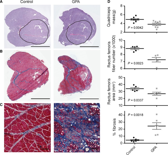
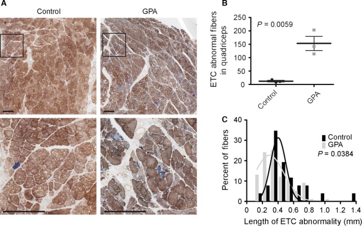
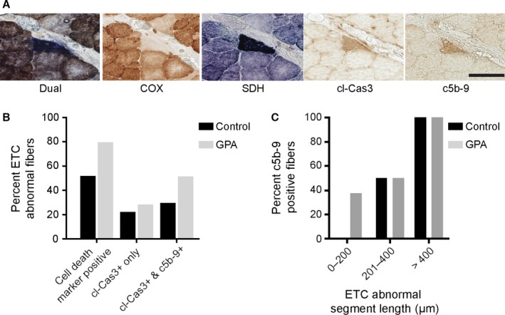
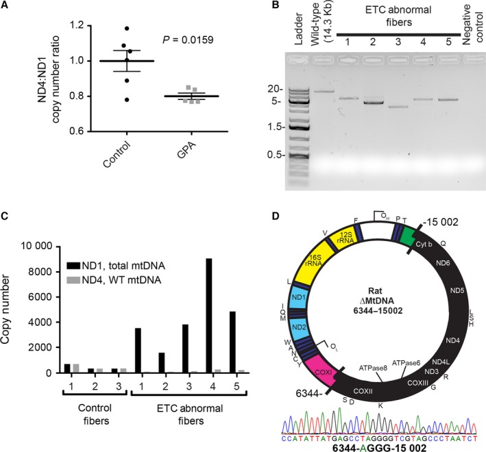
With age, somatically derived mitochondrial DNA (mtDNA) deletion mutations arise in many tissues and species. In skeletal muscle, deletion mutations clonally accumulate along the length of individual fibers. At high intrafiber abundances, these mutations disrupt individual cell respiration and are linked to the activation of apoptosis, intrafiber atrophy, breakage, and necrosis, contributing to fiber loss. This sequence of molecular and cellular events suggests a putative mechanism for the permanent loss of muscle fibers with age. To test whether mtDNA deletion mutation accumulation is a significant contributor to the fiber loss observed in aging muscle, we pharmacologically induced deletion mutation accumulation. We observed a 1200% increase in mtDNA deletion mutation-containing electron transport chain-deficient muscle fibers, an 18% decrease in muscle fiber number and 22% worsening of muscle mass loss. These data affirm the hypothesized role for mtDNA deletion mutation in the etiology of muscle fiber loss at old age.
Keywords: aging; mitochondrial DNA deletion mutation; muscle; sarcopenia.
© 2016 The Authors. Aging Cell published by the Anatomical Society and John Wiley & Sons Ltd.
Figures
References
-
- Bakeeva LE, Chentsov Yu n, Skulachev VP (1978) Mitochondrial framework (reticulum mitochondriale) in rat diaphragm muscle. Biochim. Biophys. Acta 501, 349–369. - PubMed
-
- Baris OR, Ederer S, Neuhaus JF, von Kleist‐Retzow JC, Wunderlich CM, Pal M, Wunderlich FT, Peeva V, Zsurka G, Kunz WS, Hickethier T, Bunck AC, Stockigt F, Schrickel JW, Wiesner RJ (2015) Mosaic deficiency in mitochondrial oxidative metabolism promotes cardiac arrhythmia during aging. Cell Metab. 21, 667–677. - PubMed
-
- Bergeron R, Ren JM, Cadman KS, Moore IK, Perret P, Pypaert M, Young LH, Semenkovich CF, Shulman GI (2001) Chronic activation of AMP kinase results in NRF‐1 activation and mitochondrial biogenesis. Am. J. Phys. Endocrinol. Metab. 281, E1340–E1346. - PubMed
Grants and funding
- LOF24776/Canada Foundation for Innovation
- Senior Scholar Award
- P30 AG028748/AG/NIA NIH HHS/United States
- UCSD/UCLA Diabetes Research Center Pilot and Feasibility Grant
- P30 AG028748/AG/NIA NIH HHS/United States
- UCLA Hartford Center of Excellence
- R01 AG030423/AG/NIA NIH HHS/United States
- CHE-0722519/NSF Major Research Instrumentation
- P30 DK063491/DK/NIDDK NIH HHS/United States
- CJX1-443835-WS-29646/NIH-NCRR
- Ellison Medical Foundation New Scholar Award
- American Federation for Aging Research
- Glenn Foundation for Medical Research
- K08 AG032873/AG/NIA NIH HHS/United States
LinkOut - more resources
Full Text Sources
Other Literature Sources
