. 2016 Aug 26:7:12673.
doi: 10.1038/ncomms12673.

Structural basis for oxygen degradation domain selectivity of the HIF prolyl hydroxylases

Affiliations

Structural basis for oxygen degradation domain selectivity of the HIF prolyl hydroxylases

Rasheduzzaman Chowdhury et al. Nat Commun. .

Abstract

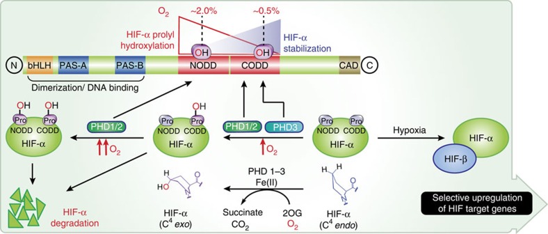
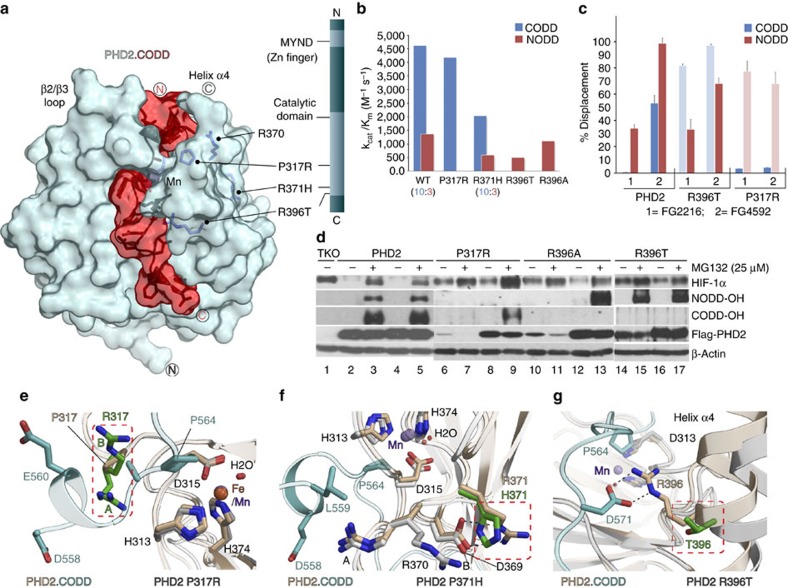
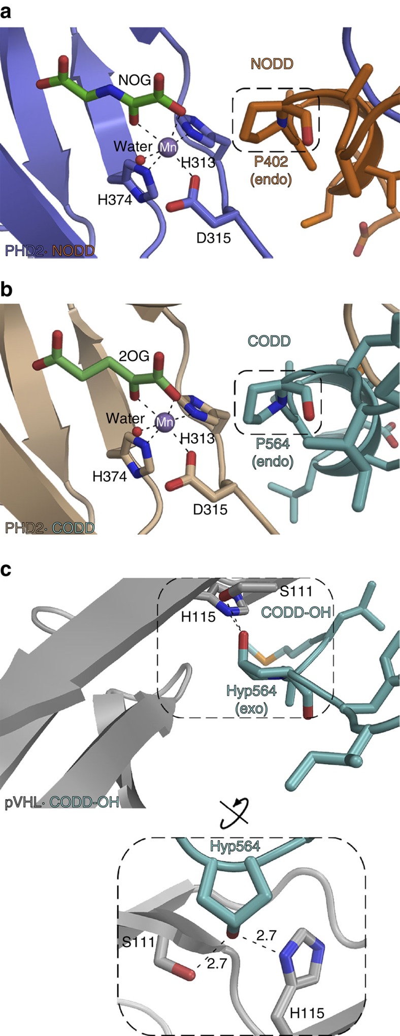
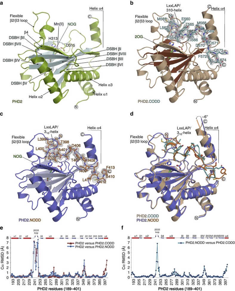
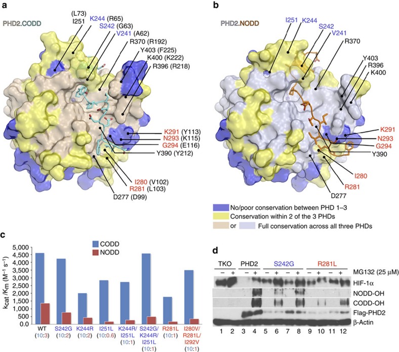
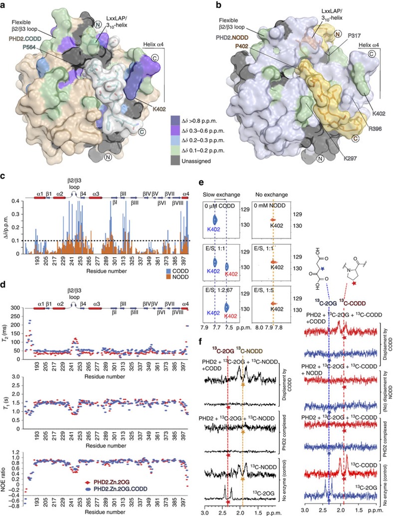
The response to hypoxia in animals involves the expression of multiple genes regulated by the αβ-hypoxia-inducible transcription factors (HIFs). The hypoxia-sensing mechanism involves oxygen limited hydroxylation of prolyl residues in the N- and C-terminal oxygen-dependent degradation domains (NODD and CODD) of HIFα isoforms, as catalysed by prolyl hydroxylases (PHD 1-3). Prolyl hydroxylation promotes binding of HIFα to the von Hippel-Lindau protein (VHL)-elongin B/C complex, thus signalling for proteosomal degradation of HIFα. We reveal that certain PHD2 variants linked to familial erythrocytosis and cancer are highly selective for CODD or NODD. Crystalline and solution state studies coupled to kinetic and cellular analyses reveal how wild-type and variant PHDs achieve ODD selectivity via different dynamic interactions involving loop and C-terminal regions. The results inform on how HIF target gene selectivity is achieved and will be of use in developing selective PHD inhibitors.


Figures

Figure 1
Figure 1. Overview of the HIF system.
The figure shows the roles of HIFα NODD/CODD hydroxylation in the hypoxic response. Ordered ODD hydroxylation is tightly regulated in animals; NODD hydroxylation is more sensitive than CODD to hypoxia. ODD hydroxylation significantly increases the affinity of hydroxylated HIFα for the VCB (VHL, elongins B and C) complex, thus signalling for HIFα degradation via proteosomal hydrolysis; the difference in kd for hydroxylated versus non-hydroxylated CODD is ∼1,000-fold (33 nM versus 34 μM, respectively). In response to hypoxia, HIFα escapes ODD hydroxylation and forms the αβ-heterodimeric HIF complex that activates the transcription of a gene array.
Figure 2
Figure 2. Clinically observed variants in PHD2 have altered selectivities.
(a) View from the PHD2.CODD complex (PDB: 3HQR) showing locations of PHD2 clinical variants with altered ODD selectivities. (b) Kinetic analyses show the P317R and R396X variants are highly selective for CODD and NODD, respectively; R371H is less efficient at the same CODD/NODD activity ratio (10:3) as wt PHD2 with (almost) unaltered selectivity. kcat/Km values are calculated from the average kcat and Km values (Supplementary Table 3). (c) One-dimensional 13C-selective clean in-phase (CLIP)–HSQC NMR reveals apparently selective displacement of NODD/CODD from PHD2 wt/clinical variant complexes by using PHD inhibitors (FG2216/ FG4592) (Supplementary Fig. 13). n=5 for wt and 2 for variants. (d) Selectivity studies using hydroxy-proline antibodies (NODD-OH and CODD-OH) and PHD 1–3 TKO MEF cells. MG 132 was used to block proteasomal degradation. In TKO cells, HIF-1α is not hydroxylated (lane 1); both NODD/CODD are fully hydroxylated in cells expressing wt PHD2 (lanes 3 and 5). Highly selective NODD/CODD hydroxylation is observed with variant PHDs irrespective of expression level of the Flag-tagged proteins. (e,f,g) Views from PHD2 P317R, R371H and R396T crystal structures superimposed with PHD2.CODD complex, suggesting substantial impact of the substitutions on substrate binding.
Figure 3
Figure 3. PHD active-site chemistry.
Binding of proline (NODD/CODD) to PHD2 and hydroxyproline/Hyp (CODD-OH) to the VHL component of the VCB complex. (a,b) Conserved binding modes of the Pro402NODD/Pro564CODD to PHD2 (PDB: 5L9V and 5L9B). It is noteworthy that the proline C4-methylene adopts the endo-conformation when bound to PHD2 in both the NODD and CODD complex structures. (c) In contrast, Hyp564CODD-OH adopts the exo- conformation when bound to the VCB complex (PDB: 1LM8). Binding of O2 is proposed to be limiting in PHD-ODD catalysis. It is noteworthy that the metal bound water, which is replaced by O2 in catalysis, is similarly positioned in both the NODD and CODD complexes (with NOG/Mn substituted for 2OG/Fe).
Figure 4
Figure 4. Overall binding modes of NODD and CODD to PHD2.
Views from PHD2.NOG (a, PDB: 5L9R), PHD2.2OG.CODD (b, PDB: 5L9B) and PHD2.NOG.NODD complexes (c, PDB: 5L9V) showing secondary structural elements involved in NODD/CODD selectivity. (d) Superimposition of PHD2.CODD and PHD2.NODD structures reveals apparently more ‘induced fit' in CODD (compared with NODD) binding involving the β2/β3 loop and α4. (e) Cα r.m.s.d. plots of PHD2 structures with/without CODD (brown) and NODD (blue) reveal similar PHD2 backbone conformations for the major and minor β-sheets of the DSBH and surrounding three α-helices (1–3), but clear differences especially in the β2/β3 loop (aa 237–250), βIV/βV loop (aa 348–353) and C-terminal helix α4 (393–401, CODD). (f) Cα r.m.s.d. plot comparing the PHD2.NODD and PHD2.CODD structures shows differences mainly in the β2/β3 loop (aa 243–250) and C-terminal regions (aa 393–401).
Figure 5
Figure 5. Combined biophysical and biochemical analyses identify NODD/CODD selectivity determinants in the PHDs.
Surface representations of (a) PHD2.CODD and (b) PHD2.NODD complexes showing sequence differences that inform on selectivity determinants in addition to clinically observed variant sites (Pro317, Arg371 and Arg396) (Supplementary Fig. 1 gives a sequence alignment of PHDs). (c) Catalytic efficiencies (kcat/Km) of PHD2 wt and the variants for hydroxylating CODD and NODD; values in the parentheses are CODD/NODD activity ratios; selected variants were tested in cells (d). Assays of the indicated PHD2 variants in TKO cells as described in Fig. 2d.
Figure 6
Figure 6. NMR studies reveal dynamics of ODD selectivity determinants in solution.
Surface representations of PHD2.CODD (CODD in grey, PDB: 5L9B) (a) and PHD2.NODD (NODD in yellow, PDB: 5L9V) (b) showing perturbed regions (colour coded) on ODD binding with differences in chemical shift values in c. On CODD binding, changes concentrate in: N terminus (α1, aa 190–195; α2, 229–231; β2/β3 loop, 234–254; β4, 255–258; and α3-βI, 294–298), the DSBH core (βI, 299–300; βII, 312–317; and βII–βIII, 319–322) and C-terminal regions including the βVIII-α4 loop (391–393) and α4 (395–402). Compared with CODD, binding of NODD induces fewer changes including in: β2/β3 loop (236–241 and 247–255), β4 (257–258), β4-α3 (259–262), α3 (268), α3-βI (290–299) βII–βIII (318–319), and βVI–βVII (369–370), and smaller changes (relative to CODD) in α4 (398–400). (d) 15N relaxation (T1/T2) and 1H-15N NOE measurements reveal β2/β3 loop dynamics that are diminished on CODD binding. (e) 15N HSQC experiments show that CODD binding directly perturbs PHD2 C terminus as exemplified by slow-exchange titration behaviour of Lys402 (291K), whereas binding of NODD has less/no influence (310K). (f) Competitive binding experiments (one-dimensional 13C-selective clean in-phase (CLIP)–HSQC) reveal CODD binding displaces NODD but not vice versa within limits of detection. 13C-2OG/ 13C-NODD/ 13C-CODD-selective excitations are indicated by coloured asterisks.

References

    1. Bishop T. & Ratcliffe P. J. HIF hydroxylase pathways in cardiovascular physiology and medicine. Circ. Res. 117, 65–79 (2015). - PMC - PubMed
    1. Bruick R. K. Oxygen sensing in the hypoxic response pathway: regulation of the hypoxia-inducible transcription factor. Genes Dev. 17, 2614–2623 (2003). - PubMed
    1. Kaelin W. G. Jr & Ratcliffe P. J. Oxygen sensing by metazoans: the central role of the HIF hydroxylase pathway. Mol. Cell 30, 393–402 (2008). - PubMed
    1. Schofield C. J. & Ratcliffe P. J. Oxygen sensing by HIF hydroxylases. Nat. Rev. Mol. Cell. Biol. 5, 343–354 (2004). - PubMed
    1. Semenza G. L. Hydroxylation of HIF-1: oxygen sensing at the molecular level. Physiology (Bethesda) 19, 176–182 (2004). - PubMed

Publication types

MeSH terms