. 2016 Sep 13;113(37):E5464-71.
doi: 10.1073/pnas.1609450113. Epub 2016 Aug 25.

Zinc-sensitive MRI contrast agent detects differential release of Zn(II) ions from the healthy vs. malignant mouse prostate

Affiliations

Zinc-sensitive MRI contrast agent detects differential release of Zn(II) ions from the healthy vs. malignant mouse prostate

M Veronica Clavijo Jordan et al. Proc Natl Acad Sci U S A. .

Abstract

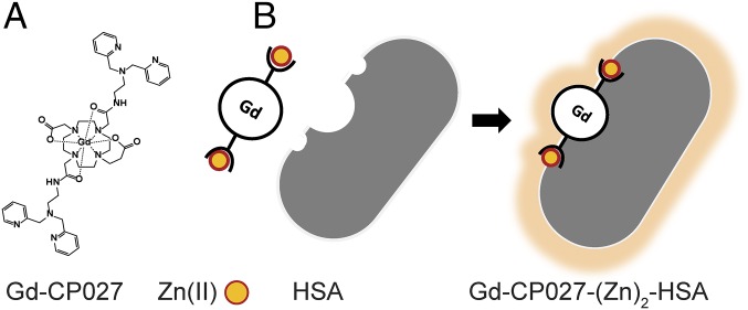
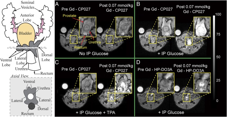
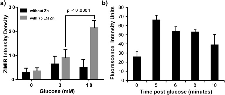
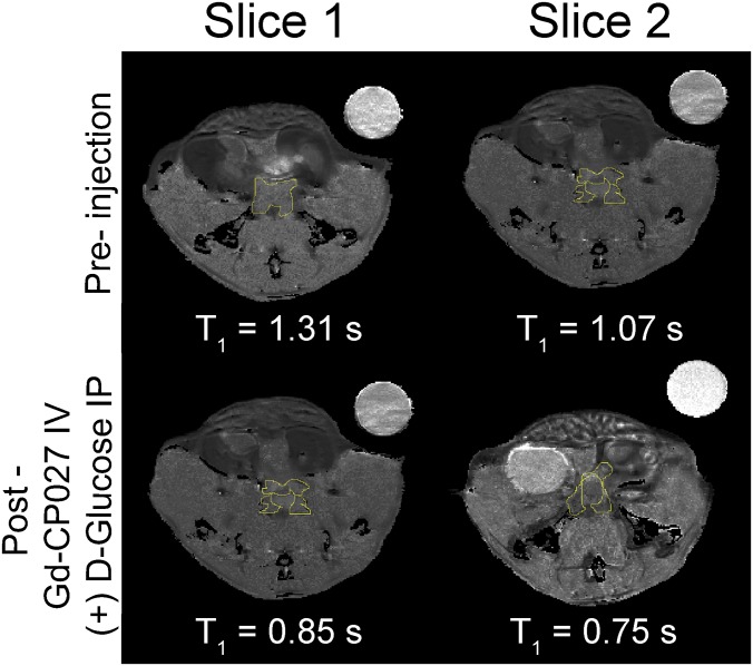
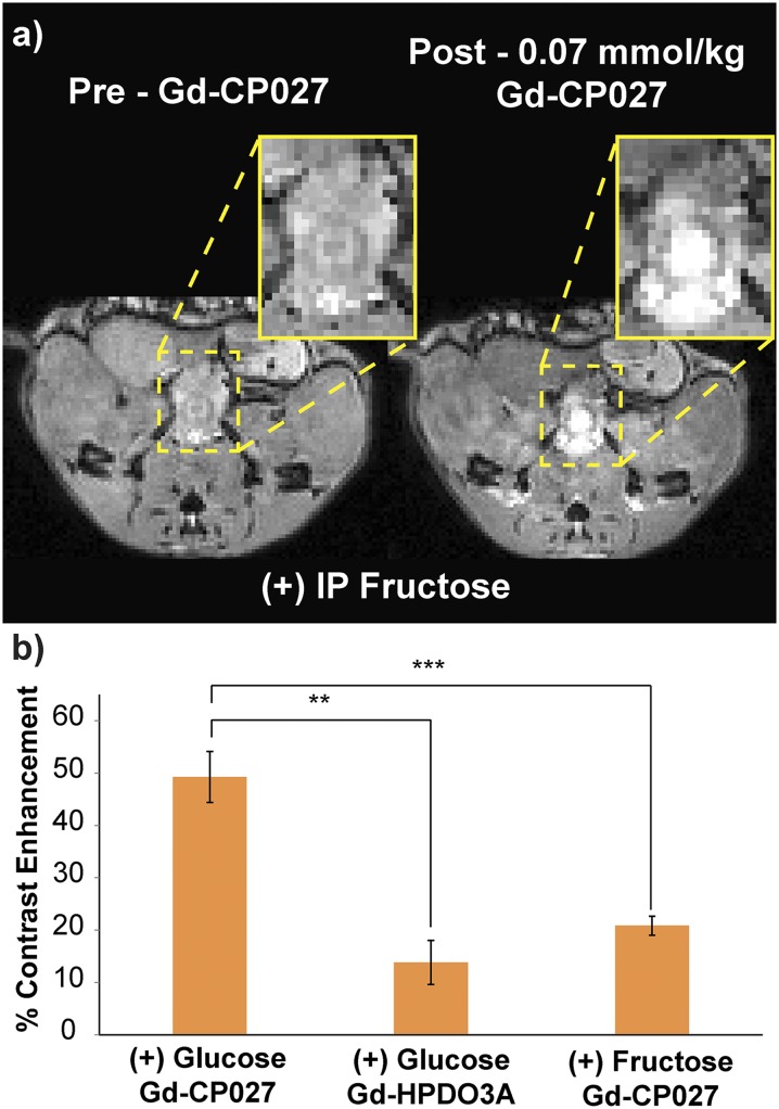
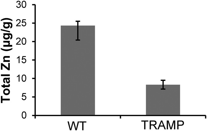
Many secretory tissues release Zn(II) ions along with other molecules in response to external stimuli. Here we demonstrate that secretion of Zn(II) ions from normal, healthy prostate tissue is stimulated by glucose in fasted mice and that release of Zn(II) can be monitored by MRI. An ∼50% increase in water proton signal enhancement is observed in T1-weighted images of the healthy mouse prostate after infusion of a Gd-based Zn(II) sensor and an i.p. bolus of glucose. Release of Zn(II) from intracellular stores was validated in human epithelial prostate cells in vitro and in surgically exposed prostate tissue in vivo using a Zn(II)-sensitive fluorescent probe known to bind to the extracellular surface of cells. Given the known differences in intracellular Zn(II) stores in healthy versus malignant prostate tissues, the Zn(II) sensor was then evaluated in a transgenic adenocarcinoma of the mouse prostate (TRAMP) model in vivo. The agent proved successful in detecting small malignant lesions as early as 11 wk of age, making this noninvasive MR imaging method potentially useful for identifying prostate cancer in situations where it may be difficult to detect using current multiparametric MRI protocols.

Keywords: MRI; cancer; glucose; prostate; zinc.


Conflict of interest statement

A.D.S., W.-H.L., and C.P. have a financial interest in VitalQuan, LLC.

Figures

Fig. 1.
Fig. 1.
Gd-based contrast agent mechanism for sensitive Zn(II) detection. (A) Gd-CP027 structure in its off configuration with a relaxivity of r1 = 5.6 mM−1⋅s−1 at B0 = 9.4 T and 6.4 mM−1⋅s−1 at B0 = 0.5 T. (B) Gd-based contrast agent binds to Zn(II) and forms a complex with human serum albumin (HSA) resulting in a change to its on configuration to form a highly sensitive MRI sensor with r1 = 9.4 mM−1⋅s−1 at B0 = 9.4 T and 48 mM−1⋅s−1 at B0 = 0.5 T.
Fig. 2.
Fig. 2.
Magnetic resonance imaging at 9.4 T of the 12 h fasted mouse prostate. Left illustrates the anatomy of an adult mouse prostate and an axial view of the gland; each visible prostatic lobe is identified in the schematic representation and can be easily correlated to the in vivo images in AD. Preinjection and postinjection of Gd-CP027 (A) without i.p. injection of d-glucose, showing no visible prostate enhancement, and (B) with simultaneous i.p. injection of d-glucose, resulting in a dramatic enhancement detected in the dorsolateral prostate as well as in the urethra. (C) IV injection of TPA 8–10 min before contrast agent. No substantial enhancement is observed in either the dorsolateral or ventral lobes. (D) Preinjection and postinjection of Gd-HPDO3A with i.p. injection of d-glucose indicating that a Zn(II) insensitive agent does not enhance prostate tissues. The urethra is intensely enhanced indicating contrast agent clearance.
Fig. S1.
Fig. S1.
Dynamic contrast measured as percent signal change in both prostate and kidney. Approximately 50% washout was observed at ∼60 min.
Fig. S2.
Fig. S2.
T1 weighted ge3d MRI (B0 = 9.4 T) of the non-Zn(II) responsive Gd-HP-DO3A injected at a dose of 0.14 mmol⋅kg−1 along with 50 µL of 20% (wt/vol) d-glucose. No significant accumulation/response to glucose in the prostate is observed. Top left circle shows water phantom used for normalization purposes.
Fig. 3.
Fig. 3.
Zn(II) secretion can be observed both in vivo and in vitro using the Zn(II)-responsive fluorescent probe, ZIMIR. (A) (Left) The two lipophilic chains on ZIMIR provide a mechanism for anchoring to the fluorescent agent to the extracellular surface of cells. (Right) In vivo experimental scheme showing placement of the microscope lens directly over the exposed prostate gland of a mouse after local administration of ZIMIR to the tissue. (B) In vivo confocal images of the prostate before (Left) and after (Right) i.p. injection of d-glucose. (C) In vitro fluorescence images of prostate epithelial cells (RWPE-1) grown in a low-glucose medium without (Left) or with (Right) 75 µM ZnSO4. The cells were then removed from the culture medium and washed with buffer, and 1 μM ZIMIR was added to coat the outer surface of the cells to detect release of Zn(II) from intracellular stores. As shown, cells not incubated with ZnSO4 did not release Zn(II) in response to glucose (Left), whereas cells that had accumulated substantial Zn(II) during incubation did release Zn(II) in response to glucose in a concentration-dependent manner.
Fig. S3.
Fig. S3.
(A) In vitro glucose-stimulated Zn(II) secretion in normal prostate epithelial cells (RWPE-1). Three different ROIs containing two cells were selected at random and measured for each treatment. Statistical significance was evaluated at a 95% confidence level. (B) In vivo confocal of exposed mouse prostate. Three ROIs were selected within the tissue field of view, ROIs placed at areas of highest intensity for each time point.
Fig. 4.
Fig. 4.
(A) Glucose-stimulated contrast enhanced (GSCE) T1-weighted 3D MR images at 9.4 T of the prostate of TRAMP mice during various stages of tumor development. (Bottom) A typical GSCE pattern of normal healthy prostate in a young TRAMP mouse (age: 10 wk). (Middle) Nascent tumor in dorsal lobe of the prostate showing clear hypointensity due to the presumable lack of intracellular Zn(II) (age: 17 wk). (Top) Poorly differentiated (PD) tumor that originated in lateral lobe and extending to ventral lobe. No GSCE was detected in the tumor, and significant loss of noncancerous prostate was observed (age: 19 wk). Intensity scale bar represents units normalized to highest image intensity as 100%. (B) Average GSCE measured over the entire prostate in three cohorts of animals. P values at the 95% confidence level for NNo lesion = 20, NWD= 5, and NPD = 2.
Fig. 5.
Fig. 5.
(A) The 3D T1-weighted image of a representative young TRAMP mouse showing no distinguishable hypointense regions in prostate after administration of glucose and Gd-CP027. H&E and PCNA immunostaining and Hematoxylin counterstain show no positive PCNA stain and only hematoxylin stained nuclei (ROI 2) correlating with normal proliferative activity. Lobes identified in MRI and H&E as the ventral lobe (VL), dorsolateral lobe (DL), and urethra (U). (B) Distinguishable hypointense region in DL after glucose and Gd-CP027 injection indicates local loss of zinc and potential malignant transformation. H&E stained section of prostate with labeled VL and DL on both sides of the urethra. PCNA of adjacent prostate section shows a spectrum of positive nuclear stain. Positive localized PCNA identified on both DLs, with one extension being predominantly more prevalent. VL shows negative PCNA stain (ROI 1) compared with evident positive stain in DL (ROI 2). (C) PD tumor stemming from lateral/ventral lobe. H&E and PCNA stains distinguish large PD tumor (T) clearly, PCNA-positive stain is observed throughout DLs of the prostate. Arrows indicate examples of cells undergoing increased proliferative activity based on positive-PCNA stain.
Fig. S4.
Fig. S4.
(A) T1-weighted MRI of a large poorly differentiated tumor bearing prostate after GSCE with Gd-CP027. Rectangle shows dorsal lobe with minimal signal enhancement. (B) H&E stain showing dorsal lobe with identified high-grade PIN areas at 20× and 40× . (C) PCNA stain of same region as in H&E showing positive PCNA indicative of malignant transformation at 20× and 40×.
Fig. S5.
Fig. S5.
T1 map of pre– and post–Gd-CP027 IV and d-glucose i.p. T1 measurements of ROIs from each slice with FLAIR pulse sequence TE/TR = 10/4,000 ms; NEX = 1; TI = 10, 50, 75, 100, 500, 750, 1,500, and 2,500 ms; and k zero = 1. Water phantom is shown in upper right corner. B0 = 9.4 T.
Fig. S6.
Fig. S6.
MR imaging of male C57bl6 mice fasted for 12 h. (A) Preinjection and postinjection of Gd-CP027 IV and 50 µL of 20% (wt/vol) fructose i.p. The dorsolateral prostate shows marginal enhancement, whereas the urethra and the rectum show the most enhancement. (B) Fructose increased contrast in the prostate up to 20%, only slightly above Gd-HPDO3A, whereas d-glucose increases contrast in the prostate up to ∼50%. **P = 0.0026, ***P = 0.0025. Error bars indicate the SEM for n = 4.
Fig. S7.
Fig. S7.
Total zinc measurements from wild-type (C57BL/6) and TRAMP mice at 25 wk of age exhibiting suspicious lesions and increased prostate volume (n = 3). A threefold decrease in zinc content was found for the TRAMP prostates compared with WT.

Similar articles

Cited by

References

    1. Lubag AJ, De Leon-Rodriguez LM, Burgess SC, Sherry AD. Noninvasive MRI of β-cell function using a Zn2+-responsive contrast agent. Proc Natl Acad Sci USA. 2011;108(45):18400–18405. - PMC - PubMed
    1. Kelleher SL, McCormick NH, Velasquez V, Lopez V. Zinc in specialized secretory tissues: Roles in the pancreas, prostate, and mammary gland. Adv Nutr. 2011;2(2):101–111. - PMC - PubMed
    1. Krebs NF. Overview of zinc absorption and excretion in the human gastrointestinal tract. J Nutr. 2000;130(5S Suppl):1374S–1377S. - PubMed
    1. Wiseman BS, et al. Site-specific inductive and inhibitory activities of MMP-2 and MMP-3 orchestrate mammary gland branching morphogenesis. J Cell Biol. 2003;162(6):1123–1133. - PMC - PubMed
    1. Seo YA, Lee S, Hennigar SR, Kelleher SL. Prolactin (PRL)-stimulated ubiquitination of ZnT2 mediates a transient increase in zinc secretion followed by ZnT2 degradation in mammary epithelial cells. J Biol Chem. 2014;289(34):23653–23661. - PMC - PubMed

Publication types