. 2016 Aug 8;1(9):16128.
doi: 10.1038/nmicrobiol.2016.128.

Structures of Ebola virus GP and sGP in complex with therapeutic antibodies

Affiliations

Structures of Ebola virus GP and sGP in complex with therapeutic antibodies

Jesper Pallesen et al. Nat Microbiol. .

Abstract

The Ebola virus (EBOV) GP gene encodes two glycoproteins. The major product is a soluble, dimeric glycoprotein (sGP) that is secreted abundantly. Despite the abundance of sGP during infection, little is known regarding its structure or functional role. A minor product, resulting from transcriptional editing, is the transmembrane-anchored, trimeric viral surface glycoprotein (GP). GP mediates attachment to and entry into host cells, and is the intended target of antibody therapeutics. Because large portions of sequence are shared between GP and sGP, it has been hypothesized that sGP may potentially subvert the immune response or may contribute to pathogenicity. In this study, we present cryo-electron microscopy structures of GP and sGP in complex with GP-specific and GP/sGP cross-reactive antibodies undergoing human clinical trials. The structure of the sGP dimer presented here, in complex with both an sGP-specific antibody and a GP/sGP cross-reactive antibody, permits us to unambiguously assign the oligomeric arrangement of sGP and compare its structure and epitope presentation to those of GP. We also provide biophysical evaluation of naturally occurring GP/sGP mutations that fall within the footprints identified by our high-resolution structures. Taken together, our data provide a detailed and more complete picture of the accessible Ebolavirus glycoprotein landscape and a structural basis to evaluate patient and vaccine antibody responses towards differently structured products of the GP gene.


Figures

Figure 1
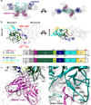
Figure 1. Structural analyses of ZMapp™ - EBOV GP complexes
a, Single particle cryo-EM reconstruction of antibodies c2G4 and c13C6 in complex with GPΔTM. There are three copies of each Fab per trimer. A single protomer from the map was segmented into c2G4 Fab (red), c13C6 Fab (blue), GP1 (cyan), and GP2 (yellow). b, Single particle cryo-EM reconstruction of antibodies c4G7 and c13C6 in complex with GPΔTM. There are two copies of each c13C6 Fab and three copies of each c4G7 Fab per trimer. A single protomer from the map was segmented into c4G7 Fab (orange), c13C6 Fab (blue), GP1 (cyan), and GP2 (yellow). c, Construct designs used for cryo-EM versus x-ray crystallography. Key differences are inclusion of the MLDs and all glycans in the proteins expressed for single particle cryo-EM. Predicted N-linked glycans are represented by magenta “Y” symbols. d, Overall structure of EBOV GP showing a side (above) and top (below) view relative to the viral membrane. Additional features not seen in the previous MLD-deleted structure include three N-linked glycans modeled at N238, N228 and N268, as well as the linker between HR1 and HR2. The putative location of the mucin-like domains, which were a part of the construct used for cryo-EM but unresolved in the maps, is indicated. SP = signal peptide (removed during processing) MPER = membrane proximal external region, TM = transmembrane region, CT = cytoplasmic tail, MLD = mucin-like domain. The TM and CT regions were not included in our constructs. Asterisks indicate glycans that were resolved in the crystal structure of EBOV GPΔMuc (PDB 3CSY).
Figure 2
Figure 2. Details of the ZMapp™ antibody epitopes
a, Epitope of antibody c2G4 bound to EBOV GPΔTM. The variable light chain is shown in dark red and the variable heavy chain in red-orange. GP2 is shown in yellow and GP1 in dark cyan. The major contacts are located at the base of the IFL and in HR1, which include residue Q508 (shown in green) and a glycan at N563. Contact regions between GP and c2G4 are resolved to ~3.9 Å (Supplementary Fig. 2). b, Epitopes and CDRs of antibody c4G7 bound to EBOV GPΔTM, with the variable light chain in dark orange and the variable heavy chain in yellow-orange. GP1 and GP2 are shown as in a). c4G7 is positioned differently from c2G4, more toward the IFL and the cathepsin loop. c4G7 also contacts part of GP1, below HR1 at the bottom of the GP chalice, in a manner similar to KZ52. Contact regions between GP and c4G7 are resolved to ~4.4 Å (Supplementary Fig. 2). c, Epitopes and CDRs of antibody c13C6 bound to EBOV GPΔTM (c2G4-c13C6 model) with variable light chain in dark blue and variable heavy chain in light blue. The major contacts are within the glycan cap, but some backbone contacts are made with the GP head. There is additional contact with glycans at N238 and N268. Contact regions between GP and c13C6 are resolved to ~4.1 Å (Supplementary Fig. 2). d, Epitopes of all three components of ZMapp™, including a comparison with antibody KZ52, which binds at the base of GP and overlaps with c2G4 and c4G7. For c13C6, contact residues are highlighted in shades of blue. For c2G4, c4G7 and KZ52, contact residues are highlighted in yellow (unique contact), orange (shared with one other base-binding mAb) or red (shared between all three base-binding mAbs). The asterisk indicates a known escape mutant for c2G4 and c4G7 at Q508. e, The GP1-GP2 interface epitope. A single protomer of the GP crystal structure PDB 3CSY is shown. The boxed area is zoomed in on the right, with mAb contact residues colored as in panel d.
Figure 3
Figure 3. Cryo-EM structure of EBOV sGP in complex with c13C6 from ZMapp™ and BDBV human survivor mAb BDBV91
a, Cryo-EM density map of EBOV sGP in complex with c13C6 and BDBV91 Fabs. sGP is in sea green (sGPA) and lime green (sGPB) with c13C6 Fabs in dark blue and BDBV91 Fabs in purple. There are two copies of each Fab bound per sGP dimer. Side (left) and top (right) views are shown. b, Subdomain assignments of sGP based on our model determined from the ~5.5 Å cryo-EM density map. A cartoon schematic of the sGP dimer is shown and corresponding domain positions are colored according to the figure below. Regions in white or black cross-hatch did not have any corresponding density, but were contained in our construct. The signal peptide (SP) in grey is excised from the construct during processing. Predicted disulfide bonds assignments are indicated between the two protomers and corresponding residues are labeled in red. Although we modeled an alanine residue for C53, we did not resolve the C53-C53 disulfide bond. c, Epitope details and CDR assignments of antibody BDBV91 bound to EBOV sGP, with the variable light chain in violet and the variable heavy chain in magenta. The sGP interface is colored as in panel b. The major contacts are formed between a single sGP protomer and CDRH3 and CDRL3. In the density map, contact regions between GP and BDBV91 (and between GP and c13C6) are resolved to ~4.4 Å (Supplementary Fig. 2). d, At hydrophobic pocket is formed at the dimer interface.and the long BDBV91 CDRH3 reaches into this pocket and contacts several residues in this region. Some side chain density between CDRH3 and sGP could be observed, including V92, K95, and F159.
Figure 4
Figure 4. Comparison of GP and sGP structures
A single protomer from sGP (left) and GP (right) is shown in the same orientation. Domains with large structural divergence are colored the same in each corresponding structure, according to sub-domain assignments in Fig. 3b. In GP, the sGP loop1 (residues 53–70, magenta) manifests as the base of the GP1 core and the C53-C609 disulfide anchoring GP1 to GP2. The hydrophobic patch in sGP (residues 179–188, lime green) forms a hydrophobic clamp on HR1 in prefusion GP, while it comprises the dimer interface in sGP. sGP loop 2 (residues 189–214, dark blue) is the cathepsin cleavage loop in GP, which reaches across the outside of GP2 on the trimer, and is processed upon entry into the endosome. The different oligomeric contexts of sGP loop 2 and the GP cathepsin cleavage loop cause these regions to adopt distinct conformations. C-terminal of W275, the structures of GP and sGP diverge. Here, sGP terminates in sGP loop 3 (yellow), which contains the C306-C306 disulfide. However, in GP, these residues form the rest of the glycan cap and the beginning of the MLD. Beyond residue 295, sGP and GP also differ in their sequences; in GP, this region becomes the bulk of the MLD (in cyan).
Figure 5
Figure 5. Effects of naturally occurring mutations that appeared in the 2014 EBOV outbreak on antibody binding within identified GP and sGP footprints
a–c, We identified several mutations in GP sequences of viruses sequenced during the 2014 EBOV outbreak that fell within the ZMapp™ footprints identified in our cryoEM reconstructions. To evaluate their effects on antibody binding, we performed site-directed mutagenesis followed by biolayer interferometry (BLI). Our BLI results were normalized to WT binding Kd values and graphed. We also included negative controls identified by alanine scanning or known escape mutants, in order to validate previous work. d–e, For sGP, we identified a single naturally occurring mutation that fell within the BDBV91 footprint (D150A), and we also identified key residues from our model to manipulate BDBV91 binding. Experiments were performed in triplicate (technical replicates, using a single preparation of GP). Error bars represent standard deviation from the mean.
Figure 6
Figure 6. Differential presentation of GP/sGP epitopes during ebolavirus infection
During infection, both sGP and GP are produced and secreted from the Golgi apparatus. While sGP is secreted in large abundance into the serum, GP is anchored to the cell membrane that forms the enveloped surface of budding virions. Antibodies detailed in this study demonstrate a portion of the antigenic landscape available during an active infection, including a GP-specific epitope at the base of GP (c2G4 and c4G7), sGP/GP cross reactive epitopes at the top of GP and sides of sGP in the glycan cap (c13C6), and an sGP-specific epitope at the sGP dimer interface (BDBV91).

Similar articles

Cited by

References

    1. Henao-Restrepo AM, et al. Efficacy and effectiveness of an rVSV-vectored vaccine expressing Ebola surface glycoprotein: interim results from the Guinea ring vaccination cluster-randomised trial. Lancet. 2015;386:857–866. - PubMed
    1. Stanley DA, et al. Chimpanzee adenovirus vaccine generates acute and durable protective immunity against ebolavirus challenge. Nature medicine. 2014;20:1126–1129. - PubMed
    1. Tapia MD, et al. Use of ChAd3-EBO-Z Ebola virus vaccine in Malian and US adults, and boosting of Malian adults with MVA-BN-Filo: a phase 1, single-blind, randomised trial, a phase 1b, open-label and double-blind, dose-escalation trial, and a nested, randomised, double-blind, placebo-controlled trial. Lancet Infect Dis. 2016;16:31–42. - PMC - PubMed
    1. Lee JE, et al. Structure of the Ebola virus glycoprotein bound to an antibody from a human survivor. Nature. 2008;454:177–182. - PMC - PubMed
    1. Lee JE, Saphire EO. Ebolavirus glycoprotein structure and mechanism of entry. Future virology. 2009;4:621–635. - PMC - PubMed

MeSH terms