. 2017 Apr 3;8(3):282-296.
doi: 10.1080/21505594.2016.1208890. Epub 2016 Aug 25.

Enterococcus faecalis readily colonizes the entire gastrointestinal tract and forms biofilms in a germ-free mouse model

Affiliations

Enterococcus faecalis readily colonizes the entire gastrointestinal tract and forms biofilms in a germ-free mouse model

Aaron M T Barnes et al. Virulence. .

Abstract

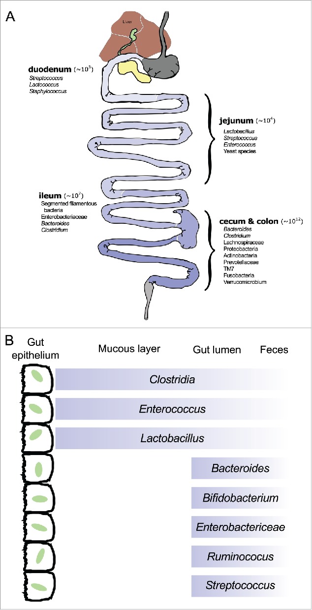
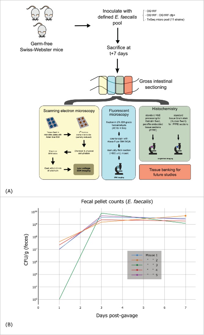
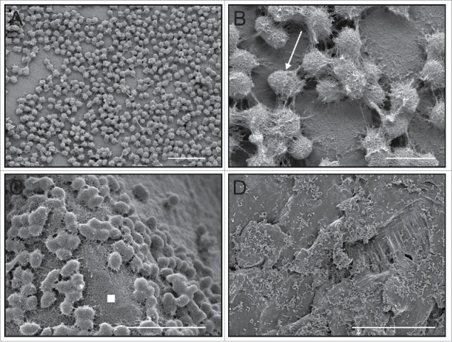
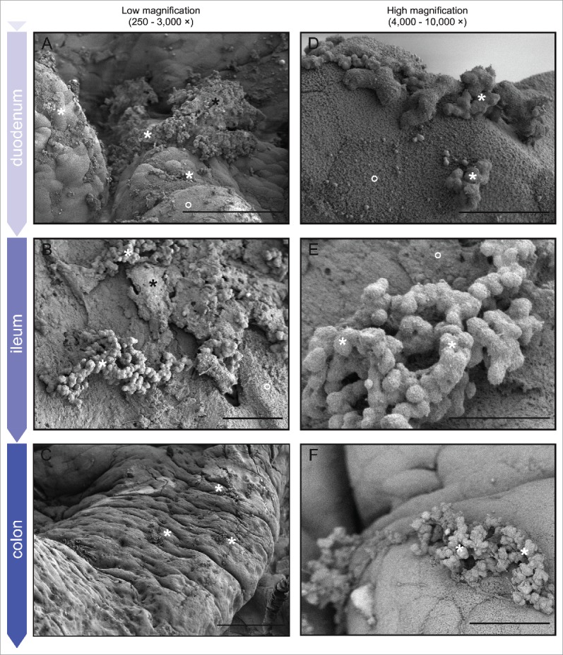
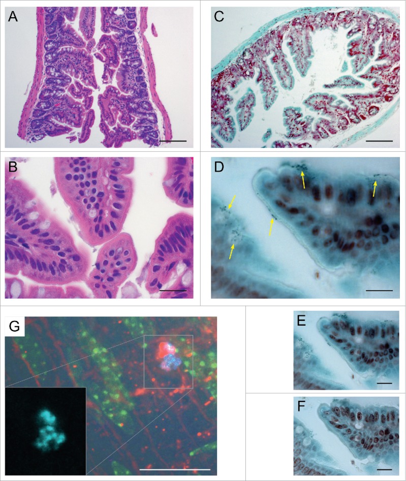
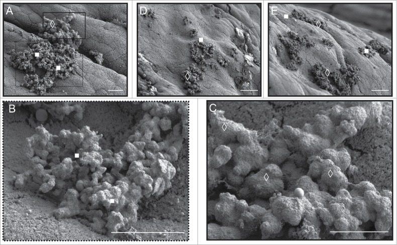
The mammalian gastrointestinal (GI) tract is a complex organ system with a twist-a significant portion of its composition is a community of microbial symbionts. The microbiota plays an increasingly appreciated role in many clinically-relevant conditions. It is important to understand the details of biofilm development in the GI tract since bacteria in this state not only use biofilms to improve colonization, biofilm bacteria often exhibit high levels of resistance to common, clinically relevant antibacterial drugs. Here we examine the initial colonization of the germ-free murine GI tract by Enterococcus faecalis-one of the first bacterial colonizers of the naïve mammalian gut. We demonstrate strong morphological similarities to our previous in vitro E. faecalis biofilm microcolony architecture using 3 complementary imaging techniques: conventional tissue Gram stain, immunofluorescent imaging (IFM) of constitutive fluorescent protein reporter expression, and low-voltage scanning electron microscopy (LV-SEM). E. faecalis biofilm microcolonies were readily identifiable throughout the entire lower GI tract, from the duodenum to the colon. Notably, biofilm development appeared to occur as discrete microcolonies directly attached to the epithelial surface rather than confluent sheets of cells throughout the GI tract even in the presence of high (>109) fecal bacterial loads. An in vivo competition experiment using a pool of 11 select E. faecalis mutant strains containing sequence-defined transposon insertions showed the potential of this model to identify genetic factors involved in E. faecalis colonization of the murine GI tract.

Keywords: antibiotic resistance; competitive fitness; intestinal microbiota; opportunistic pathogen.


Figures

Figure 1.
Figure 1.
(A) Anatomy of the murine gastrointestinal tract with general mammalian microbial spatial distribution pattern. Values in parentheses indicate average total bacterial cells per gram of host tissue. (B) Cross-section of murine GI tract demonstrating localization of bacterial species relative to the gut epithelium (after Sekirov et al. 2010).
Figure 2.
Figure 2.
(A) Experimental overview of sample preparation and processing. (B) Time series of E. faecalis recovered from fecal samples of initially germ free mice after inoculation by oral gavage (CFUs are per g of recovered feces).
Figure 3.
Figure 3.
Four previously-unpublished low-voltage scanning electron microscopy (LV-SEM) of E. faecalis microcolony and biofilm formation under a range of on in vitro conditions similar to our previously published work demonstrating both the rapidity in which attachment and extracellular matrix production begins as well as the general structural morphology of the ECM when the matrix is properly preserved using a cationic dye stabilization system. (A) Aclar fluoropolymer coupons, 24 hrs, CDC biofilm reactor system, bar = 6 µm;51 (B) higher magnification from (A) showing the E. faecalis diplococci surrounded by the sweater-like ECM (arrow), bar = 1 µm; (C) note that the biofilm ECM not only covers cells, but extends into interstitial space between cell clusters (white filled square), regenerated cellulose membrane, 24 hrs, bar = 5 µm;50 (D) polycarbonate coupon, 24 hrs, bar = 50 µm.
Figure 4.
Figure 4.
LV-SEM of representative murine GI tract tissue sacrificed 7 d post-gavage with E. faecalis. Rows show samples from each of the major divisions of the lower GI tract from the proximal duodenum through the distal colon; columns show increasing micrograph magnification (left to right). For space reasons low magnification micrographs demonstrating anatomic structure and micrographs of surface colonization in the jejunum and cecum are presented separately in Fig S1. White stars = E. faecalis microcolonies; black stars = retained fecal material; open circles = mucinous material (host). Left column: The traditional biofilm ECM structure of E. faecalis microcolonies can be seen throughout the lower GI tract. Note the relatively large distances between many microcolonies even 7 d post-inoculation (mostly clearly in C).Right column: Biofilm microcolonies exhibit wide size variation, from a few tens of cells (D) to several hundred or more (E). Note also the generally bland nature of the underlying host epithelium (D, F)Bars: A = 50 µm; B, C, F = 10 µm, D = 1 mm, E = 200 µm.
Figure 5.
Figure 5.
Histological and IFM examination of murine GI tissue from representative sections adjacent to those prepared for SEM.A-F: Histology of the colonized murine terminal ileum. (A). H&E demonstrating normal ileal morphology; note marked lack of obvious inflammatory response. (B). Higher magnification of A. (C). Tissue Gram stain (Hucker-Twort) at low magnification. (D). Higher magnification of C showing numerous small, Gram-positive organisms (yellow arrows showing clusters of small, dark blue cells at the luminal border of epithelial cells. (E). Additional image of same section ∼1 μm deeper in the tissue. (F). Additional image of same section ∼2 μm deeper where the murine epithelium is in focus. G: IFM of the colonized proximal murine colon. (G). Immunofluorescent microscopy of the murine proximal colon colonized by the constitutive CFP-expressing strain OG1RF cfp+. Blue (cyan) = CFP-positive E. faecalis cells; red = Alexa Fluor 594. WGA ; green = autofluorescence from the murine epithelium. The red WGA lectin antibody primarily labels the host epithelial layer as well, but some labeling can be seen around bacterial cells that have begun to produce polysaccharide-rich extracellular matrix. Bars: A, C = 100 µm; B, D - F = 20 µm.
Figure 6.
Figure 6.
Morphological variation in bacterial extracellular matrix apparently specific to biofilm microcolonies in mice gavaged with a small, TnSeq-derived mutant pool of 11 E. faecalis. (A) E. faecalis microcolonies attached to murine colonic epithelium. Under higher magnification, the foreground cluster (white filled squares; higher magnification in B) appears to have a rougher, less voluminous extracellular matrix compared to the background microcolony ECM (white open diamonds; higher magnification in C). Additional examples of other areas of the colon exhibit the same duality (D, E). Murine colon colonized by the parental OG1RF E. faecalis strain only appears to form the classic ECM form predominantly seen in (C) – compare with Figure 4E. Bars: A - C = 5 µm; D, E = 30 µm.

Similar articles

Cited by

References

    1. Sekirov I, Russell SL, Antunes LCM, Finlay BB. Gut microbiota in health and disease. Physiol Rev [Internet] 2010. [cited 2014December17]; 90:859-904. Available from: http://www.ncbi.nlm.nih.gov/pubmed/20664075; PMID:20664075; http://dx.doi.org/10.1152/physrev.00045.2009 - DOI - PubMed
    1. Quigley EMM. Small intestinal bacterial overgrowth: what it is and what it is not. Curr Opin Gastroenterol [Internet] 2014. [cited 2016January4]; 30:141-6. Available from: http://www.ncbi.nlm.nih.gov/pubmed/24406476; PMID:24406476; http://dx.doi.org/10.1097/MOG.0000000000000040 - DOI - PubMed
    1. Berg RD. The indigenous gastrointestinal microflora. Trends Microbiol [Internet] 1996. [cited 2016January7]; 4:430-5. Available from: http://www.ncbi.nlm.nih.gov/pubmed/8950812; PMID:8950812; http://dx.doi.org/10.1016/0966-842X(96)10057-3 - DOI - PubMed
    1. Savage DC, Dubos R, Schaedler RW. The gastrointestinal epithelium and its autochthonous bacterial flora. J Exp Med [Internet] 1968. [cited 2015December31]; 127:67-76. Available from: http://www.pubmedcentral.nih.gov/articlerender.fcgi?artid=2138434&tool=p...; PMID:4169441; http://dx.doi.org/10.1084/jem.127.1.67 - DOI - PMC - PubMed
    1. Brown EM, Sadarangani M, Finlay BB. The role of the immune system in governing host-microbe interactions in the intestine. Nat Immunol [Internet] 2013. [cited 2014September15]; 14:660-7. Available from: http://www.ncbi.nlm.nih.gov/pubmed/23778793; PMID:23778793; http://dx.doi.org/10.1038/ni.2611 - DOI - PubMed

Publication types

MeSH terms

LinkOut - more resources