

Feline Calcium Oxalate Urolithiasis: Risk factors and rational treatment approaches
- PMID: 27562981
- PMCID: PMC11148900
- DOI: 10.1177/1098612X16660442
Feline Calcium Oxalate Urolithiasis: Risk factors and rational treatment approaches
Abstract
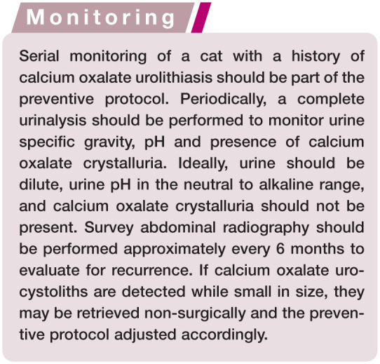
Practical relevance: Uroliths occur commonly in the bladder and/or urethra of cats and can be lifethreatening if urethral obstruction occurs. Calcium oxalate accounts for 40-50% of urocystoliths and these stones are not amenable to medical dissolution; therefore, removal by surgery or minimally invasive techniques is required if uroliths must be treated. Medical protocols for prevention involve decreasing urine saturation for minerals that form uroliths.
Etiopathogenesis: Formation of uroliths is not a disease, but rather a complication of several disorders. Some disorders can be identified and corrected (such as infection-induced struvite urolith formation); others can be identified but not corrected (such as idiopathic hypercalcemia). In most cats with calcium oxalate urolith formation the underlying etiopathogenesis is not known. A common denominator of all these disorders is that they can from time to time create oversaturation of urine with one or more crystal precursors, resulting in formation of crystals.
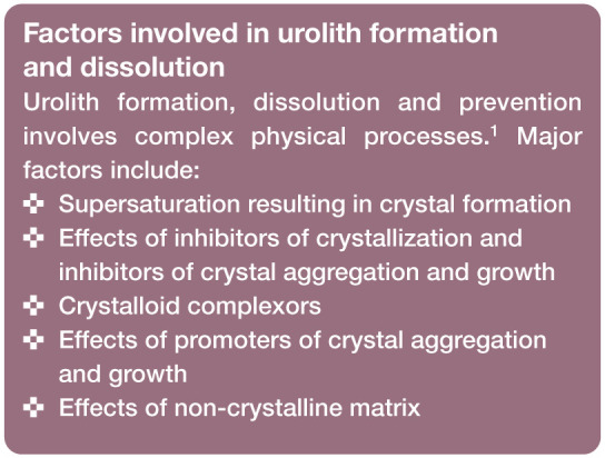
Basic concepts: In order to develop rational and effective approaches to treatment, abnormalities that promote urolith formation must be identified, with the goal of eliminating or modifying them. It is important, therefore, to understand several basic concepts associated with urolithiasis and the factors that promote urolith formation that may be modified with medical treatment; for example, the state of urinary saturation, modifiers of crystal formation, potential for multiple crystal types, and presence of bacterial infection or urinary obstruction.
© The Author(s) 2016.
Conflict of interest statement
The author declared no potential conflicts of interest with respect to the research, authorship and/or publication of this article.
Figures
References
-
- Bartges JW, Osborne CA, Lulich JP, et al.. Methods for evaluating treatment of uroliths. Vet Clin North Am Small Anim Pract 1999; 29: 45–57. - PubMed
-
- Feeney DA, Anderson KL. Radiographic imaging in urinary tract disease. In: Bartges J, Polzin DJ. (eds). Nephrology and urology of small animals. Ames, IA: Wiley-Blackwell, 2011, pp 97–127.
-
- Osborne CA, Lulich JP, Kruger JM, et al.. Analysis of 451,891 canine uroliths, feline uroliths, and feline urethral plugs from 1981 to 2007: perspectives from the Minnesota Urolith Center. Vet Clin North Am Small Anim Pract 2009; 39: 183–197. - PubMed
-
- Langston C, Gisselman K, Palma D, et al.. Diagnosis of urolithiasis. Compend Contin Educ Pract Vet 2008; 30: 447–450, 452–444; quiz 455. - PubMed
Publication types
MeSH terms
Substances
LinkOut - more resources
Full Text Sources
Other Literature Sources
Miscellaneous
