Review
. 2016 Sep 15;10(5):687-98.
doi: 10.5009/gnl16033.

Biology of Cholangiocytes: From Bench to Bedside

Affiliations
Review

Biology of Cholangiocytes: From Bench to Bedside

Kyo-Sang Yoo et al. Gut Liver. .

Abstract

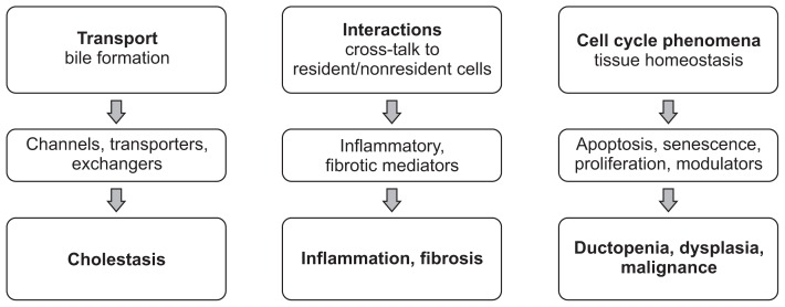
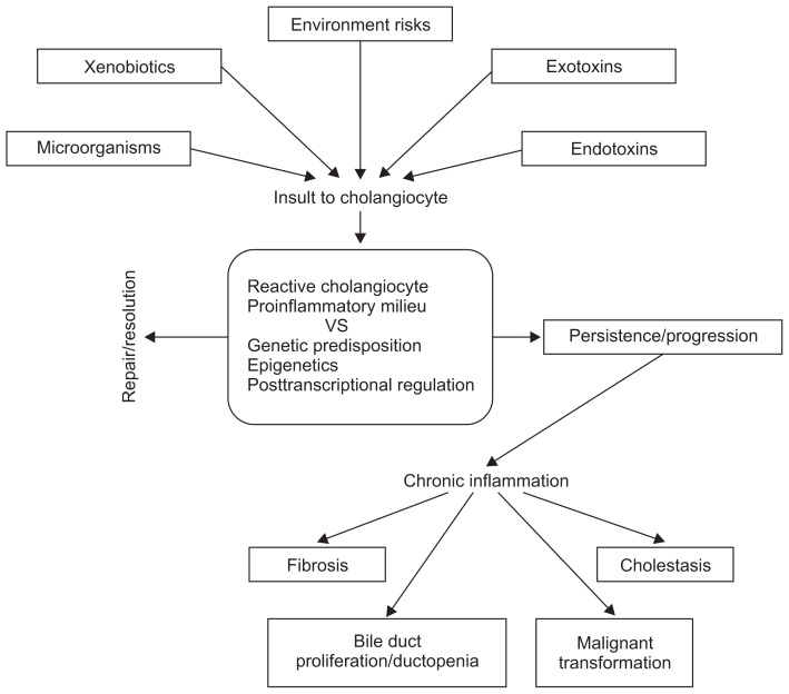
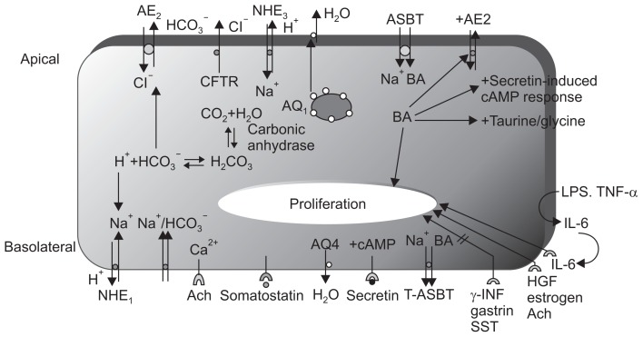
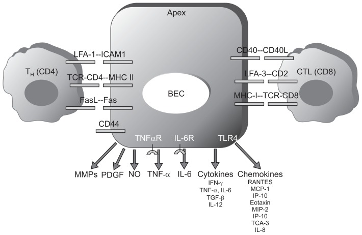
Cholangiocytes, the lining epithelial cells in bile ducts, are an important subset of liver cells. They are activated by endogenous and exogenous stimuli and are involved in the modification of bile volume and composition. They are also involved in damaging and repairing the liver. Cholangiocytes have many functions including bile production. They are also involved in transport processes that regulate the volume and composition of bile. Cholangiocytes undergo proliferation and cell death under a variety of conditions. Cholangiocytes have functional and morphological heterogenecity. The immunobiology of cholangiocytes is important, particularly for understanding biliary disease. Secretion of different proinflammatory mediators, cytokines, and chemokines suggests the major role that cholangiocytes play in inflammatory reactions. Furthermore, paracrine secretion of growth factors and peptides mediates extensive cross-talk with other liver cells, including hepatocytes, stellate cells, stem cells, subepithelial myofibroblasts, endothelial cells, and inflammatory cells. Cholangiopathy refers to a category of chronic liver diseases whose primary disease target is the cholangiocyte. Cholangiopathy usually results in end-stage liver disease requiring liver transplant. We summarize the biology of cholangiocytes and redefine the concept of cholangiopathy. We also discuss the recent progress that has been made in understanding the pathogenesis of cholangiopathy and how such progress has influenced therapy.

Keywords: Biology; Cholangiocyte; Cholangiopathy; Receptors; Transport.


Figures

Fig. 1
Fig. 1
Biology of cholangiocytes.,,, Various molecules conduct several important functions in cholangiocytes. Bile is formed through the activity of transmembrane molecules, such as channels, transporters, and exchangers. Dysfunction of these molecules may lead to cholestasis. Cholangiocytes interact with resident and nonresident cells of bile ducts via inflammatory and fibrotic mediators, such as tumor necrosis factor α and interleukin 6, which, in disease states, results in biliary inflammation and fibrosis. Cholangiocytes contribute to the cell-cycle phenomena that maintain tissue homeostasis via modulators of apoptosis, senescence, and proliferation. In disease states, these processes may result in ductopenia, dysplasia, and malignant transformation of the bile ducts.
Fig. 2
Fig. 2
Pathogenic model of cholangiopathy.,,, Cholangiocytes interact with endogenous or exogenous substances, microorganisms, or environmental factors. The initial host response is the development of a reactive cholangiocyte and a proinflammatory microenvironment. The balance of the host response to insult depends on genetic susceptibility, epigenetics, and posttranscriptional regulation, and it may result in the resolution of the disease state or the perpetuation of the initial inflammatory response. This may result in chronic inflammation of the bile ducts and ultimately to cholestasis, bile duct proliferation, ductopenia, fibrosis, and the potential malignant transformation of cholangiocytes.
Fig. 3
Fig. 3
Membrane transporters/receptors/channel proteins in cholangiocytes.,– The choleretic effect of secretin is mediated by increases in cAMP, the activation of cAMP-dependent Cl channels (CFTR), and the subsequent ductal secretion of bicarbonate. Aquaporin water channels (regulated by secretin) appear to play an important role in this process. cAMP-dependent Cl secretion enables the mobilization of intracellular Ca2+ stores and the activation of Ca2+-dependent apical membrane Cl channels. The NHE1 isoform regulates secretin-stimulated ductal secretion. Numerous hormone/peptide receptors have been identified on the basolateral domain of cholangiocytes. Several of these receptors (VIP and bombesin) modulate ductal choleresis, whereas other receptors (gastrin and somatostatin) inhibit basal and secretin-stimulated choleresis. The apically located ABAT enables the entry of bile salts into cholangiocytes, whereas the truncated form of ABAT eliminates bile salts from the basolateral membrane. AE, anion exchanger; CFTR, cystic fibrosis transmembrane conductance regulator; NHE, sodium-hydrogen exchanger; ASBT, apical sodium-dependent bile acid transporter; cAMP, cyclic adenosin mono-phpsphate; LPS, lipopolysaccharide; TNF, tumor necrosis factor; IL, interleukin; HGF, hepatocyte growth factor; Ach, acetylcholine; INF, interferon; SST, somatostatin; VIP, vasoactive intestinal peptide; ABAT, apical bile acid transporter.
Fig. 4
Fig. 4
Immune properties of cholangiocytes. Cholangiocytes express adhesion molecules, which enable their interaction with CD4+ and CD8+ T cells. Because of the expression of major histocompatibility complex (MHC)-I and MHC-II on their surface, cholangiocytes are cytotoxic targets and/or antigen-presenting cells (APCs). Cholangiocytes produce chemokines and cytokines, which have autocrine or paracrine effects and modulate immune reactions. In addition, cholangiocytes secrete metalloproteinases, nitric oxide, and other growth factors involved in immune injury and fibrogenesis of the liver. LFA, lymphocyte function-associated antigen; TCR, T-cell receptor; BEC, biliary epithelial cell; CTL, cytotoxic T lymphocyte; MMP, matrix metal-loproteinase; PDGF, platelet-derived growth factor; NO, nitric oxide; TNF, tumor necrosis factor; IL, interleukin; IFN, interferon; TGF, transforming growth factor; MCP, monocyte chemotactic protein; IP-10, interferon-inducible protein-10; MIP-2, macrophage inflammatory protein-2; TCA, T-cell activation gene-3.

References

    1. Alpini G, McGill JM, Larusso NF. The pathobiology of biliary epithelia. Hepatology. 2002;35:1256–1268. doi: 10.1053/jhep.2002.33541. - DOI - PubMed
    1. Strazzabosco M, Spirlí C, Okolicsanyi L. Pathophysiology of the intrahepatic biliary epithelium. J Gastroenterol Hepatol. 2000;15:244–253. doi: 10.1046/j.1440-1746.2000.02091.x. - DOI - PubMed
    1. Roberts SK, Ludwig J, Larusso NF. The pathobiology of biliary epithelia. Gastroenterology. 1997;112:269–279. doi: 10.1016/S0016-5085(97)70244-0. - DOI - PubMed
    1. Tietz PS, Larusso NF. Cholangiocyte biology. Curr Opin Gastroenterol. 2006;22:279–287. doi: 10.1097/01.mog.0000218965.78558.bc. - DOI - PubMed
    1. Strazzabosco M, Fabris L, Spirli C. Pathophysiology of cholangiopathies. J Clin Gastroenterol. 2005;39(4 Suppl 2):S90–S102. doi: 10.1097/01.mcg.0000155549.29643.ad. - DOI - PubMed