. 2016 Sep 30;44(17):8013-9.
doi: 10.1093/nar/gkw708. Epub 2016 Aug 25.

3.9 Å structure of the nucleosome core particle determined by phase-plate cryo-EM

Affiliations

3.9 Å structure of the nucleosome core particle determined by phase-plate cryo-EM

Eugene Y D Chua et al. Nucleic Acids Res. .

Abstract

The Volta phase plate is a recently developed electron cryo-microscopy (cryo-EM) device that enables contrast enhancement of biological samples. Here we have evaluated the potential of combining phase-plate imaging and single particle analysis to determine the structure of a small protein-DNA complex. To test the method, we made use of a 200 kDa Nucleosome Core Particle (NCP) reconstituted with 601 DNA for which a high-resolution X-ray crystal structure is known. We find that the phase plate provides a significant contrast enhancement that permits individual NCPs and DNA to be clearly identified in amorphous ice. The refined structure from 26,060 particles has an overall resolution of 3.9 Å and the density map exhibits structural features consistent with the estimated resolution, including clear density for amino acid side chains and DNA features such as the phosphate backbone. Our results demonstrate that phase-plate cryo-EM promises to become an important method to determine novel near-atomic resolution structures of small and challenging samples, such as nucleosomes in complex with nucleosome-binding factors.


Figures

Figure 1.
Figure 1.
Phase-plate cryo-EM analysis of Nucleosome Core Particles (NCPs) in ice. (A and C) Field views of NCP particles recorded with the Volta phase plate. Representative views of the NCP circled in blue (disk view), in red (tilted view) and in green (side view). Arrows indicate excess DNA, in white (bent DNA) and in black (straight DNA). Scale bars are 50 nm. (B) Examples of reference-free 2D class averages showing representative disk, tilted and side views. Mask diameter is 12 nm.
Figure 2.
Figure 2.
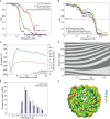
Data processing and map statistics. (A) Fourier Shell Correlation (FSC) plots for the EM density map. Arrows indicate 3.9 Å resolution estimate at FSC=0.143. A comparison of the FSC corrected (red), masked (orange), unmasked (green) and phase-randomized (purple) map is shown. (B) FSC of the EM map and atomic models derived from the X-ray structure (purple) and the refined model (dark blue) of the NCP, DNA alone (orange) and protein alone (green). (C) Per-movie-frame B-factors and Guinier plot intercepts. (D) Frequency-dependent weighting of movie frames (frame numbers in maroon). The contributions of early and late frames are down-weighted due to particle movement and beam damage. (E-F) Local resolution estimate in ResMap showing that the protein density is better refined as compared to the DNA density (also shown in B).
Figure 3.
Figure 3.
3.9 Å phase-plate cryo-EM structure of the Nucleosome Core Particle (NCP). (A) Three views of the EM map (surface rendered in grey) and the refined EM model (cartoon representation). The disk view (top) and the tilted view (middle) is related by a 60° rotation about the vertical axis. The disk view (top) and the side view (lower panel) is related by a 90° rotation about the vertical axis. The DNA is shown in blue and histone octamer in pink. (B) Close-up views of histone helices and the DNA dyad. Also shown is a comparison of the EM model (gold) and the X-ray model (green; PDB ID 3LZ0) displaying a similar arrangement of the histone core and small differences in DNA conformations at the entry/exit site of the NCP.

References

    1. Bai X.C., McMullan G., Scheres S.H. How cryo-EM is revolutionizing structural biology. Trends Biochem. Sci. 2015;40:49–57. - PubMed
    1. Cheng Y., Grigorieff N., Penczek P.A., Walz T. A primer to single-particle cryo-electron microscopy. Cell. 2015;161:438–449. - PMC - PubMed
    1. Nogales E., Scheres S.H. Cryo-EM: A unique tool for the visualization of macromolecular complexity. Mol. Cell. 2015;58:677–689. - PMC - PubMed
    1. Henderson R. The potential and limitations of neutrons, electrons and X-rays for atomic resolution microscopy of unstained biological molecules. Q. Rev. Biophys. 1995;28:171–193. - PubMed
    1. Baker L.A., Rubinstein J.L. Radiation damage in electron cryomicroscopy. Methods Enzymol. 2010;481:371–388. - PubMed

Publication types