. 2016 Oct 21;291(43):22357-22372.
doi: 10.1074/jbc.M116.752626. Epub 2016 Aug 25.

Targeted Disruption of the Interaction between WD-40 Repeat Protein 5 (WDR5) and Mixed Lineage Leukemia (MLL)/SET1 Family Proteins Specifically Inhibits MLL1 and SETd1A Methyltransferase Complexes

Affiliations

Targeted Disruption of the Interaction between WD-40 Repeat Protein 5 (WDR5) and Mixed Lineage Leukemia (MLL)/SET1 Family Proteins Specifically Inhibits MLL1 and SETd1A Methyltransferase Complexes

Nilda L Alicea-Velázquez et al. J Biol Chem. .

Abstract

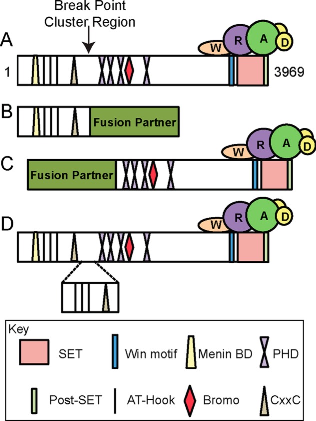
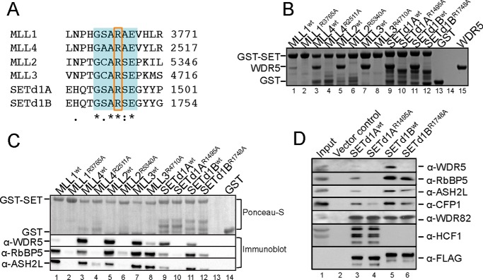
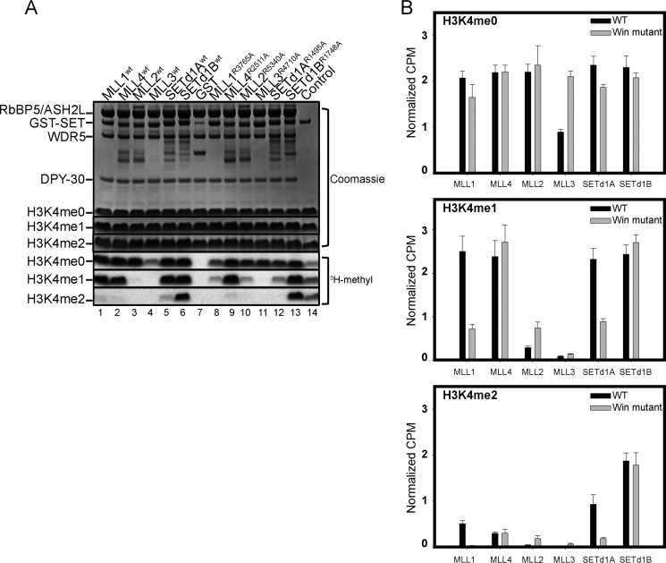
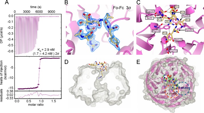
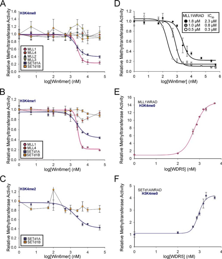
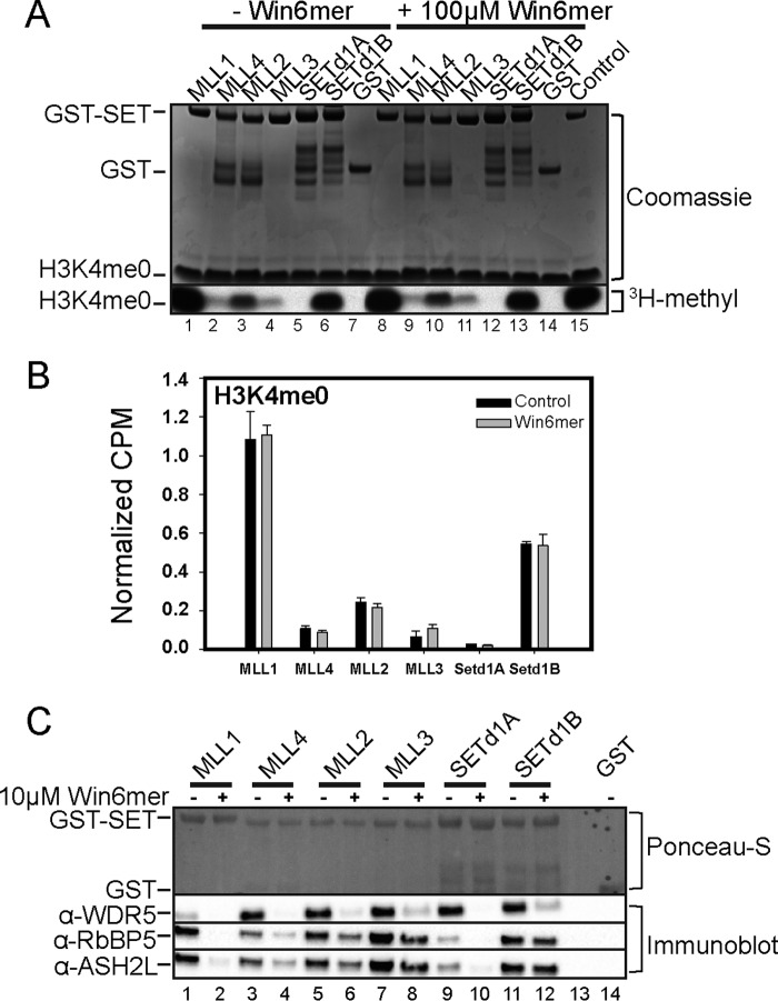
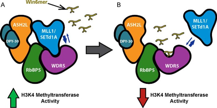
MLL1 belongs to the SET1 family of histone H3 lysine 4 (H3K4) methyltransferases, composed of MLL1-4 and SETd1A/B. MLL1 translocations are present in acute leukemias, and mutations in several family members are associated with cancer and developmental disorders. MLL1 associates with a subcomplex containing WDR5, RbBP5, ASH2L, and DPY-30 (WRAD), forming the MLL1 core complex required for H3K4 mono- and dimethylation and transcriptional activation. Core complex assembly requires interaction of WDR5 with the MLL1 Win (WDR5 interaction) motif, which is conserved across the SET1 family. Agents that mimic the SET1 family Win motif inhibit the MLL1 core complex and have become an attractive approach for targeting MLL1 in cancers. Like MLL1, other SET1 family members interact with WRAD, but the roles of the Win motif in complex assembly and enzymatic activity remain unexplored. Here, we show that the Win motif is necessary for interaction of WDR5 with all members of the human SET1 family. Mutation of the Win motif-WDR5 interface severely disrupts assembly and activity of MLL1 and SETd1A complexes but only modestly disrupts MLL2/4 and SETd1B complexes without significantly altering enzymatic activity in vitro Notably, in the absence of WDR5, MLL3 interacts with RAD and shows enhanced activity. To further probe the role of the Win motif-WDR5 interaction, we designed a peptidomimetic that binds WDR5 (Kd ∼3 nm) and selectively inhibits activity of MLL1 and SETd1A core complexes within the SET1 family. Our results reveal that SET1 family complexes with the weakest Win motif-WDR5 interaction are more susceptible to Win motif-based inhibitors.

Keywords: MLL1; SETd1A; WDR5; Win motif; enzyme; epigenetics; histone methylation; protein structure; protein-protein interaction.


Figures

FIGURE 1.
FIGURE 1.
Schematic of common outcomes of MLL1 genetic rearrangements. A, domain map of full-length wild type MLL1 with the breakpoint region denoted by an arrow. MLL1 contains many domains involved in binding chromatin (i.e. AT-Hooks, CxxC domains, PHD, and bromodomains) as well as domains involved in mediating protein-protein interactions (i.e. menin binding domain (Menin BD) and the WDR5 interaction (Win) motif). The SET and post-SET domains are involved in catalysis. The WRAD subcomplex interacts with the C terminus of MLL1. B, the most common outcome of MLL1 genetic rearrangements, which result in replacement of the C terminus of MLL1 with one of ∼70 known fusion partners (i.e. AF9, AF4, and ENL). In this arrangement, the N terminus is retained, but the catalytic SET/post-SET domain is lost. C, a rearrangement that results in the replacement of the N terminus of MLL1 with a fusion partner (i.e. AF4). In this arrangement, the N-terminal domains are lost, but the catalytic domain is retained. D, result of a partial tandem duplication in which a segment of the N terminus (containing the AT-Hooks and CxxC domain) is duplicated and inserted at the break point region.
FIGURE 2.
FIGURE 2.
The Win motif is required for interaction with WDR5. A, sequence alignment of the human SET1 family Win motif generated using Clustal Omega (90). The 6-residue Win motif is highlighted in blue, and the conserved arginine is enclosed in a box. B, GST pull-down of wild type and Win mutant SET1 family members with WDR5. Individual GST-tagged SET domains (wild type or mutant) were incubated with purified WDR5 and glutathione-coated agarose beads. A Coomassie Blue-stained SDS-polyacrylamide gel of the pull-down fractions only is shown. GST was used as a negative control (lane 13), and a sample of purified WDR5 was run on the gel to compare the migration of pull-down bands (lane 15). C, comparison of wild type or mutant GST-SET domain pull-down of WDR5, RbBP5, and ASH2L from MCF-7 cell extracts. WRA components were detected by Western blotting. The top panel shows a Ponceau S-stained PVDF membrane, and the bottom panels show the immunoblots. D, comparison of wild type or Win motif mutant FLAG-tagged full-length SETd1A and SETd1B co-immunoprecipitation with WDR5, RbBP5, ASH2L, CFP1, WDR82, and HCF1 from stably transfected T-REx HEK293 cells.
FIGURE 3.
FIGURE 3.
Substitution of the Win motif arginine alters SET1 core complex-catalyzed H3K4 methyltransferase activity in vitro. A, sample gel showing the comparison of core complex methyltransferase activities among SET1 family members (wild type and mutant) in the presence of WRAD. The top panels show Coomassie Blue-stained SDS-polyacrylamide gels, and the bottom panels show 3H-methyl incorporation after 4 h of exposure as detected by fluorography. The control lane shows the activity of the MLL1WT SET domain with 100 μm H3K4me0 peptide, which is included on each gel. B, quantification of radioactivity from excised histone H3 bands by LSC. Data are normalized to the activity level of the control lane on each gel. Error bars, S.E. of measurement among three independent experiments.
FIGURE 4.
FIGURE 4.
Co-crystal structure of Win6mer and WDR5 at 2.0 Å resolution. A, thermodynamic characterization of the WDR5-Win6mer interaction by ITC. The binding affinity Kd is reported with a confidence interval of 2σ or 95%. B, simulated annealing FoFc omit map contoured at 3σ unambiguously shows electron density corresponding to the Win6mer peptide. C, intra-Win6mer and Win6mer-WDR5 bonding network. Hydrogen bonds are represented by dashed lines. Participating residues are labeled in dark yellow (Win6mer) and purple (WDR5). Position P0 corresponds to the conserved arginine residue. Positions P− and P+ correspond to residues N- and C-terminal of P0, respectively. D, cut-away of a surface rendition of the Win6mer/WDR5 structure. WDR5 is shown in gray, and the Win6mer peptide is shown in yellow. The conserved arginine is inserted into the Win motif binding pocket in WDR5. E, overlay of the MLL1 Win motif peptide (green) (Protein Data Bank code 3EG6 (56)) and Win6mer peptide (yellow). The conserved arginines are oriented in a highly similar manner within the central cavity in WDR5 (magenta). The P+4 residue in both peptides binds the A-pocket (orange dashed circle) and not the B-pocket (blue dashed pocket) (49).
FIGURE 5.
FIGURE 5.
Inhibition of SET1 family core complex activity by Win6mer. The activity of SET1 family core complexes upon titration of Win6mer was assessed via scintillation proximity assay. SET1 family core complexes were assayed according to their substrate specificity as follows (16). A, all SET1 core complexes were assayed for monomethylation (H3K4me0 substrate). B, MLL1, MLL4, SETd1A, and SETd1B core complexes were assayed for dimethylation (H3K4me1 substrate). C, SETd1A and SETd1B core complexes were assayed for trimethylation (H3K4me2 substrate). IC50 values are reported on Table 3. D, efficiency of MLL1 core complex inhibition by Win6mer is dependent on enzyme concentration. IC50 values are shown in the inset. Activity data for each SET1 family member were normalized to the activity of uninhibited core complex. Data were fit to a dose response with variable slope equation (Equation 1). Shown is monomethyltransferase activity of MLL1-RAD (E) and SETd1A-RAD (F) upon titration of WDR5. Activity data were normalized to the activity of MLL1-RAD or SETd1A-RAD in the absence of WDR5. Data were fit to a dose response with variable slope equation (Equation 1). Error bars, S.E. of measurement between two independent experiments.
FIGURE 6.
FIGURE 6.
The Win6mer peptide interferes with core complex assembly for a subset of SET1 family members. A, representative gel of a comparison of monomethyltransferase activities among isolated SET1 family SET domains in the absence of WRAD treated with (100 μm) or without Win6mer. The top panel shows the Coomassie Blue-stained SDS-polyacrylamide gel, and the bottom panel shows 3H-methyl incorporation into H3K4 peptide after 4 h of exposure, as detected by fluorography. The control lane shows the activity of the MLL1WT SET domain on 100 μm H3K4me0 peptide. B, quantification of radioactivity from excised histone H3 peptide bands by LSC. Data are normalized to the activity level of the control lane on each gel. Error bars, S.E. of measurement among three independent experiments. C, comparison of core complex assembly by SET1 family members from pull-down experiments from MCF-7 cell extracts in the presence (+) (10 μm) or absence (−) of Win6mer. Individual GST-tagged SET domains were incubated with cell extracts and pulled down with glutathione-agarose beads. WRA components were detected by Western blotting. The top panel shows a Ponceau S-stained PVDF membrane, and the bottom panels show the immunoblots. GST (not treated with Win6mer) was used as a negative control (lane 14).
FIGURE 7.
FIGURE 7.
Model for down-regulation of MLL1 and SETd1A core complex activity by Win6mer. A, MLL1 and SETd1A SET domains require interaction with WDR5 for stabilizing assembly with the components of RAD. Like all SET1 family members, MLL1 and SETd1A utilize the Win motif to interact with WDR5. Stably assembled MLL1 and SETd1A core complexes exhibit full H3K4 methyltransferase activity. B, treatment with Win6mer, a Win motif peptidomimetic, competes with MLL1 and SETd1A SET domains for WDR5 binding, thus destabilizing core complex assembly. This, in turn, leads to down-regulation of MLL1 and SETd1A core complex activity.

References

    1. Lee J. H., Tate C. M., You J. S., and Skalnik D. G. (2007) Identification and characterization of the human Set1B histone H3-Lys4 methyltransferase complex. J. Biol. Chem. 282, 13419–13428 - PubMed
    1. Lee J. H., and Skalnik D. G. (2005) CpG-binding protein (CXXC finger protein 1) is a component of the mammalian Set1 histone H3-Lys4 methyltransferase complex, the analogue of the yeast Set1/COMPASS complex. J. Biol. Chem. 280, 41725–41731 - PubMed
    1. Ruault M., Brun M. E., Ventura M., Roizès G., and De Sario A. (2002) MLL3, a new human member of the TRX/MLL gene family, maps to 7q36, a chromosome region frequently deleted in myeloid leukaemia. Gene 284, 73–81 - PubMed
    1. Huntsman D. G., Chin S. F., Muleris M., Batley S. J., Collins V. P., Wiedemann L. M., Aparicio S., and Caldas C. (1999) MLL2, the second human homolog of the Drosophila trithorax gene, maps to 19q13.1 and is amplified in solid tumor cell lines. Oncogene 18, 7975–7984 - PubMed
    1. Prasad R., Zhadanov A. B., Sedkov Y., Bullrich F., Druck T., Rallapalli R., Yano T., Alder H., Croce C. M., Huebner K., Mazo A., and Canaani E. (1997) Structure and expression pattern of human ALR, a novel gene with strong homology to ALL-1 involved in acute leukemia and to Drosophila trithorax. Oncogene 15, 549–560 - PubMed

MeSH terms

Substances