

Angiotensin II type I receptor (AT1R) is an independent prognosticator of esophageal squamous cell carcinoma and promotes cells proliferation via mTOR activation
- PMID: 27564102
- PMCID: PMC5341864
- DOI: 10.18632/oncotarget.11567
Angiotensin II type I receptor (AT1R) is an independent prognosticator of esophageal squamous cell carcinoma and promotes cells proliferation via mTOR activation
Abstract
Background: The aim of this study was to investigate the effects of the angiotensin II/ angiotensin II type I receptor (AT1R) and angiotensin II type II receptor (AT2R) signaling pathway in esophageal squamous cell carcinoma (ESCC).
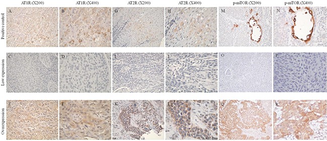
Methods: Immunohistochemistry was performed to evaluate the expression levels of AT1R and AT2R in tissues from 152 surgically resected ESCC patients, and those expression levels were then correlated with treatment outcomes. The angiotensin II/AT1R/AT2R signaling pathway and its biological effects in the context of ESCC were investigated in vitro and in vivo.
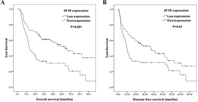
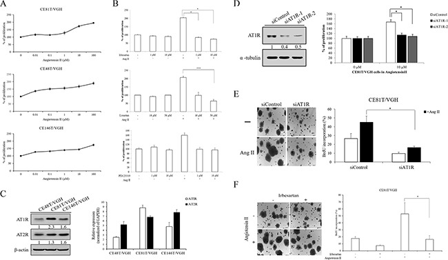
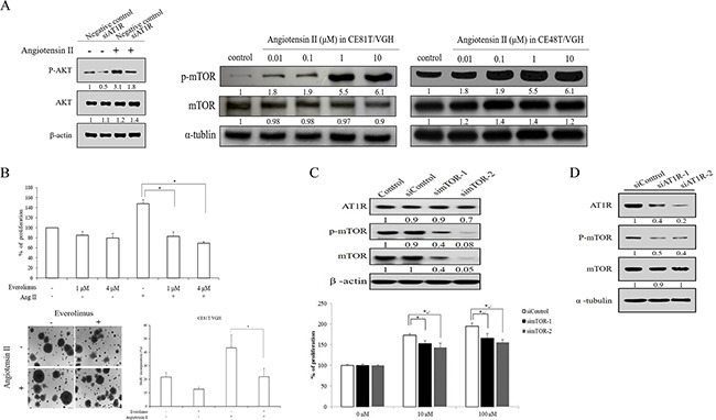
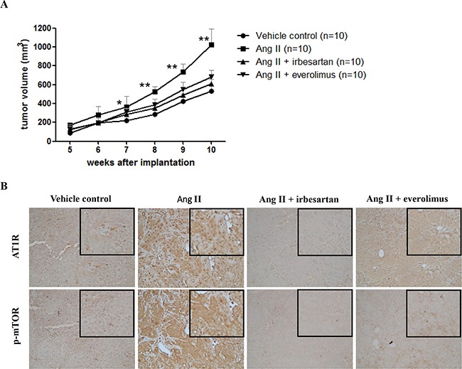
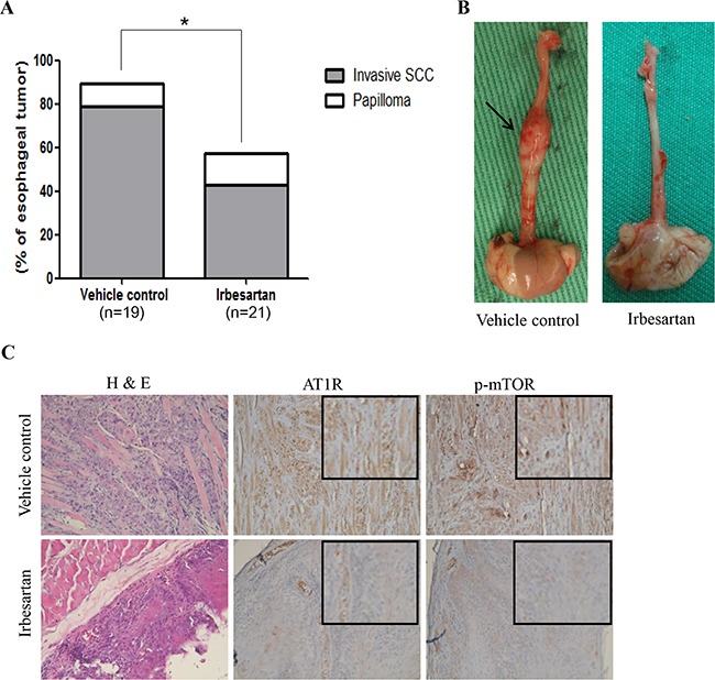
Results: In human samples, AT1R overexpression was univariately associated with inferior overall survival and remained multivariately independent (hazard ratio=1.812). In vitro, angiotensin II stimulated the growth of ESCC cells in a dose-dependent manner. Treatment with irbesartan or AT1R-RNAi knockdown but not treatment with PD123319 significantly decreased the level of angiotensin II-induced ESCC cell proliferation. Angiotensin II also caused mTOR activation in a dose-dependent manner, and everolimus or mTOR-RNAi knockdown significantly suppressed the level of angiotensin II-induced ESCC cell proliferation. Furthermore, AT1R-RNAi knockdown suppressed the activation of mTOR. Clinically, AT1R expression was also correlated with phosphorylated mTOR expression. In a xenograft model, local angiotensin II injection enhanced tumor growth, and this effect could be decreased by treatment with irbesartan or everolimus. In a 4-NQO-induced-ESCC murine model, irbesartan significantly decreased the incidence of esophageal tumor.
Conclusions: These findings suggest that AT1R overexpression is an independent adverse prognosticator for patients with ESCC and that angiotensin II/AT1R signaling stimulates ESCC growth, in part through mTOR activation.
Keywords: AT1R; angiotensin II; esophageal cancer; mTOR; squamous cell carcinoma.
Conflict of interest statement
The authors declare no conflict of interest.
Figures
Similar articles
-
Phosphorylated p70S6K expression is an independent prognosticator for patients with esophageal squamous cell carcinoma.Surgery. 2015 Mar;157(3):570-80. doi: 10.1016/j.surg.2014.10.014. Epub 2014 Nov 6. Surgery. 2015. PMID: 25726316
-
CXCL12 expression promotes esophageal squamous cell carcinoma proliferation and worsens the prognosis.BMC Cancer. 2016 Jul 21;16:514. doi: 10.1186/s12885-016-2555-z. BMC Cancer. 2016. PMID: 27439769 Free PMC article.
-
SOX2 promotes tumor growth of esophageal squamous cell carcinoma through the AKT/mammalian target of rapamycin complex 1 signaling pathway.Cancer Sci. 2013 Jul;104(7):810-6. doi: 10.1111/cas.12155. Epub 2013 Apr 16. Cancer Sci. 2013. PMID: 23510069 Free PMC article.
-
Chloride intracellular channel 1 promotes esophageal squamous cell carcinoma proliferation via mTOR signalling.Transl Oncol. 2023 Jan;27:101560. doi: 10.1016/j.tranon.2022.101560. Epub 2022 Oct 14. Transl Oncol. 2023. PMID: 36252281 Free PMC article. Review.
-
Tumor xenograft animal models for esophageal squamous cell carcinoma.J Biomed Sci. 2018 Aug 29;25(1):66. doi: 10.1186/s12929-018-0468-7. J Biomed Sci. 2018. PMID: 30157855 Free PMC article. Review.
Cited by
-
AGTR1 potentiates the chemotherapeutic efficacy of cisplatin in esophageal carcinoma through elevation of intracellular Ca2+ and induction of apoptosis.Int J Oncol. 2025 Apr;66(4):32. doi: 10.3892/ijo.2025.5738. Epub 2025 Mar 14. Int J Oncol. 2025. PMID: 40084687 Free PMC article.
-
Telmisartan Induces Osteosarcoma Cells Growth Inhibition and Apoptosis Via Suppressing mTOR Pathway.Open Life Sci. 2018 Jul 5;13:242-249. doi: 10.1515/biol-2018-0029. eCollection 2018 Jan. Open Life Sci. 2018. PMID: 33817089 Free PMC article.
-
Platelet-to-lymphocyte ratio is an independent prognosticator in patients with esophageal squamous cell carcinoma receiving esophagectomy.J Thorac Dis. 2019 Nov;11(11):4583-4590. doi: 10.21037/jtd.2019.11.06. J Thorac Dis. 2019. PMID: 31903247 Free PMC article.
-
The renin-angiotensin-aldosterone system (RAAS) signaling pathways and cancer: foes versus allies.Cancer Cell Int. 2023 Oct 27;23(1):254. doi: 10.1186/s12935-023-03080-9. Cancer Cell Int. 2023. PMID: 37891636 Free PMC article. Review.
-
Elucidating the Association Between the Upregulation of Angiotensin Type 1-Receptors and the Development of Gastrointestinal Malignancies.J Gastrointest Cancer. 2021 Jun;52(2):399-406. doi: 10.1007/s12029-020-00547-0. Epub 2020 Nov 10. J Gastrointest Cancer. 2021. PMID: 33174118 Review.
References
-
- National Department of Health. Taiwan, Republic of China Cancer Registry Annual Report 1972-2011
-
- Li SH, Rau KM, Lu HI, Wang YM, Tien WY, Liang JL, Lin WC. Pre-treatment maximal oesophageal wall thickness is independently associated with response to chemoradiotherapy in patients with T3-4 oesophageal squamous cell carcinoma. Eur J Cardiothorac Surg. 2012 - PubMed
-
- Lee PC, Mirza FM, Port JL, Stiles BM, Paul S, Christos P, Altorki NK. Predictors of recurrence and disease-free survival in patients with completely resected esophageal carcinoma. J Thorac Cardiovasc Surg. 2011;141:1196–1206. - PubMed
-
- Arrieta O, Pineda-Olvera B, Guevara-Salazar P, Hernandez-Pedro N, Morales-Espinosa D, Ceron-Lizarraga TL, Gonzalez-De la Rosa CH, Rembao D, Segura-Pacheco B, Sotelo J. Expression of AT1 and AT2 angiotensin receptors in astrocytomas is associated with poor prognosis. Br J Cancer. 2008;99:160–166. - PMC - PubMed
MeSH terms
Substances
LinkOut - more resources
Full Text Sources
Other Literature Sources
Medical
Miscellaneous
