. 2016 Aug 26;13(1):203.
doi: 10.1186/s12974-016-0687-3.

Etifoxine improves sensorimotor deficits and reduces glial activation, neuronal degeneration, and neuroinflammation in a rat model of traumatic brain injury

Affiliations

Etifoxine improves sensorimotor deficits and reduces glial activation, neuronal degeneration, and neuroinflammation in a rat model of traumatic brain injury

Emmanuelle Simon-O'Brien et al. J Neuroinflammation. .

Abstract

Background: Traumatic brain injury (TBI) results in important neurological impairments which occur through a cascade of deleterious physiological events over time. There are currently no effective treatments to prevent these consequences. TBI is followed not only by an inflammatory response but also by a profound reorganization of the GABAergic system and a dysregulation of translocator protein 18 kDa (TSPO). Etifoxine is an anxiolytic compound that belongs to the benzoxazine family. It potentiates GABAergic neurotransmission, either through a positive allosteric effect or indirectly, involving the activation of TSPO that leads to an increase in neurosteroids synthesis. In several models of peripheral nerve injury, etifoxine has been demonstrated to display potent regenerative and anti-inflammatory properties and to promote functional recovery. Prior study also showed etifoxine efficacy in reducing brain edema in rats. In light of these positive results, we used a rat model of TBI to explore etifoxine treatment effects in a central nervous system injury, from functional outcomes to the underlying mechanisms.

Methods: Male Sprague-Dawley rats received contusion (n = 18) or sham (n = 19) injuries centered laterally to bregma over the left sensorimotor cortex. They were treated with etifoxine (50 mg/kg, i.p.) or its vehicle 30 min following injury and every day during 7 days. Rats underwent behavioral testing to assess sensorimotor function. In another experiment, injured rats (n = 10) or sham rats (n = 10) received etifoxine (EFX) (50 mg/kg, i.p.) or its vehicle 30 min post-surgery. Brains were then dissected for analysis of neuroinflammation markers, glial activation, and neuronal degeneration.

Results: Brain-injured rats exhibited significant sensorimotor function deficits compared to sham-injured rats in the bilateral tactile adhesive removal test, the beam walking test, and the limb-use asymmetry test. After 2 days of etifoxine treatment, behavioral impairments were significantly reduced. Etifoxine treatment reduced pro-inflammatory cytokines levels without affecting anti-inflammatory cytokines levels in injured rats, reduced macrophages and glial activation, and reduced neuronal degeneration.

Conclusions: Our results showed that post-injury treatment with etifoxine improved functional recovery and reduced neuroinflammation in a rat model of TBI. These findings suggest that etifoxine may have a therapeutic potential in the treatment of TBI.

Keywords: Astrogliosis; Cytokines; Etifoxine; Functional recovery; Microglia; Neuroinflammation; Neuronal degeneration; Neurosteroids; TSPO; Traumatic brain injury.


Figures
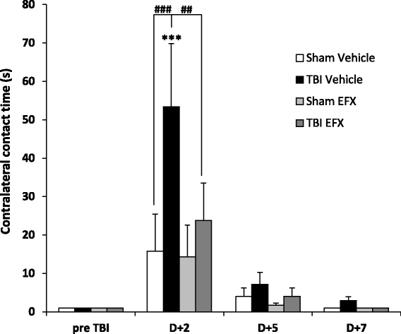
Fig. 1
Fig. 1
Bilateral adhesive removal test. Mean (+SEM) time to contact the contralateral adhesive in sham-vehicle (n = 9), TBI-vehicle (n = 10), sham-EFX (n = 10), and TBI-EFX (n = 8) groups. EFX treatment significantly improved the time to contact on day 2 post-CCI compared with vehicle treatment in TBI animals (## p < 0.01). ***p < 0.001 compared to pre-TBI; ### p < 0.001 and ## p < 0.01 compared to TBI-vehicle
Fig. 2
Fig. 2
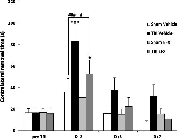
Bilateral adhesive removal test. Mean (+SEM) time to remove the contralateral adhesive in sham-vehicle (n = 9), TBI-vehicle (n = 10), sham-EFX (n = 10), and TBI-EFX (n = 8) groups. EFX treatment significantly improved the time to remove the adhesive on day 2 post-CCI compared with vehicle treatment in TBI animals (# p < 0.05). ***p < 0.001 and *p < 0.05 compared to pre-TBI; ### p < 0.001 and # p < 0.05 compared to TBI-vehicle
Fig. 3
Fig. 3
Tapered beam walking test. Mean (+SEM) percentage of faults with the contralateral hind limb on the beam in sham-vehicle (n = 9), TBI-vehicle (n = 10), sham-EFX (n = 10), and TBI-EFX (n = 8) groups. Treatment with EFX significantly improved motor performance and coordination on day 2 post-CCI compared to vehicle treatment in TBI animals (# p < 0.05). ***p < 0.001 compared to pre-TBI; ### p < 0.001 and # p < 0.05 compared to TBI-vehicle
Fig. 4
Fig. 4
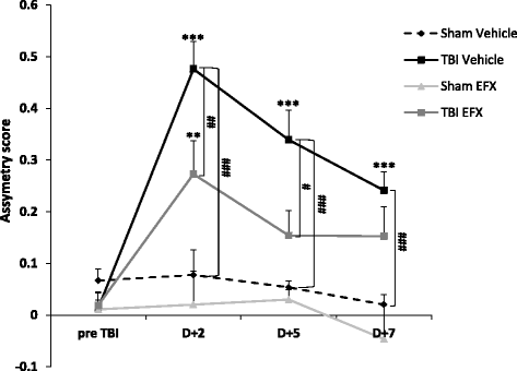
Limb-use asymmetry test. Mean (+SEM) asymmetry score in sham-vehicle (n = 9), TBI-vehicle (n = 10), sham-EFX (n = 10), and TBI-EFX (n = 8) groups. Higher scores indicate a greater use of the unimpaired forelimb. Treatment with EFX significantly improved asymmetry score on day 2 (## p < 0.01) and day 5 (# p < 0.05) post-CCI compared to vehicle treatment in TBI animals. ***p < 0.001 and **p < 0.01 compared to pre-TBI; ### p < 0.001, ## p < 0.01, and # p < 0.05 compared to TBI-vehicle
Fig. 5
Fig. 5
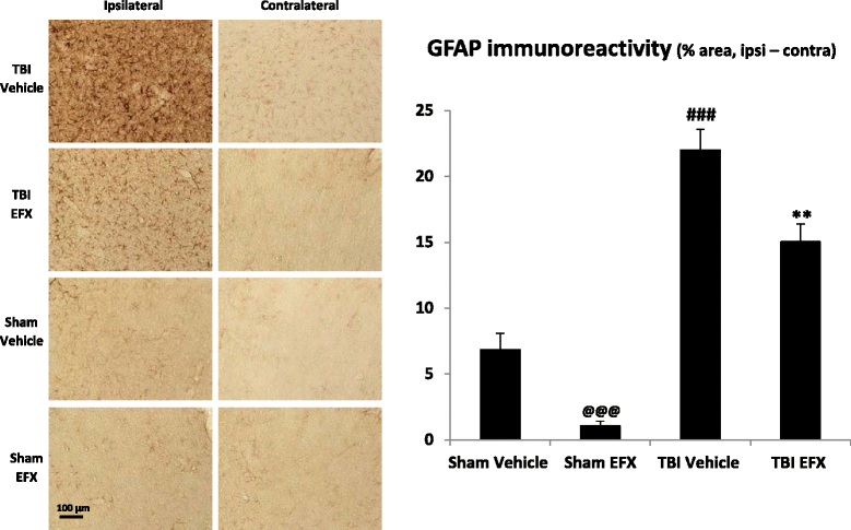
Effect of EFX treatment on CCI-induced astrogliosis in rats 2 days post-injury. EFX (50 mg/kg, i.p.) or its vehicle were administered 30 min after surgery and 24 h after (n = 5/group). Astrogliosis was evaluated with GFAP immunostaining. TBI induced an increase in GFAP immunoreactivity in the ipsilateral side of the injury (p < 0.001). EFX treatment significantly reduced astrogliosis (p < 0.01) in injured animals in comparison with vehicle treatment. Images show representative sections from each group (ipsilateral and contralateral side). Data are mean + SEM. @@@ p < 0.001: comparison between sham-vehicle and sham-EFX groups; ### p < 0.001: comparison between sham-vehicle and TBI-vehicle groups; **p < 0.01: comparison between TBI-vehicle and TBI-EFX groups
Fig. 6
Fig. 6
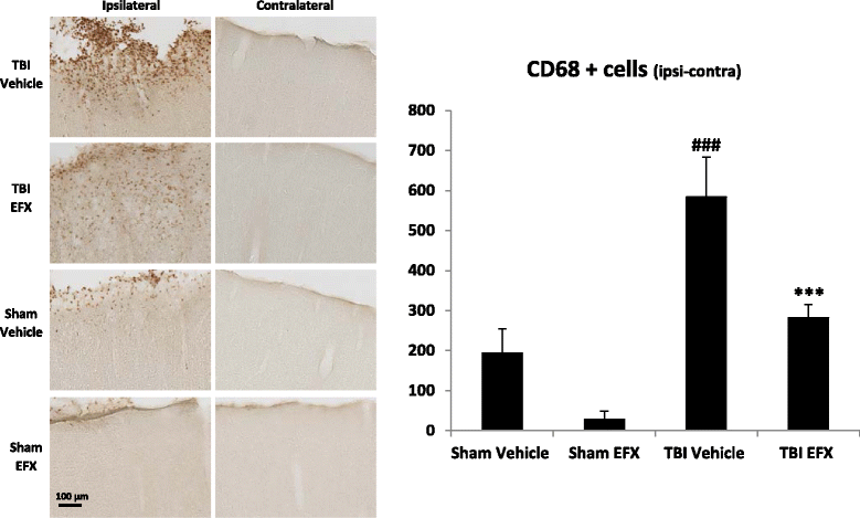
Effect of EFX treatment on CCI-induced microglia/macrophage activation in rats 2 days post-injury. EFX (50 mg/kg, i.p.) or its vehicle were administered 30 min after surgery and 24 h after (n = 5/group). Microglia/macrophage activation was evaluated with CD68 immunostaining. TBI induced an increase in CD68 immunoreactivity in the ipsilateral side of the injury (p < 0.001). EFX treatment significantly reduced microglia/macrophage activation (p < 0.001) in injured animals in comparison with vehicle treatment. Images show representative sections from each group (ipsilateral and contralateral side). Data are mean + SEM. ### p < 0.001: comparison between sham-vehicle and TBI-vehicle groups; ***p < 0.001: comparison between TBI-vehicle and TBI-EFX groups
Fig. 7
Fig. 7
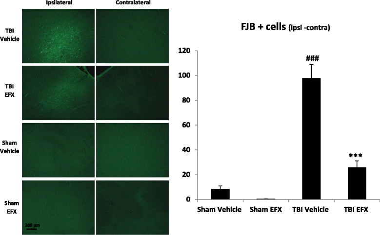
Effect of EFX treatment on CCI-induced neuronal degeneration in rats 2 days post-injury. EFX (50 mg/kg, i.p.) or its vehicle were administered 30 min after surgery and 24 h after (n = 5/group). Neuronal degeneration was evaluated with fluoro-jade B (FJB) staining. TBI induced an increase in FJB staining in the ipsilateral side of the injury (p < 0.001). EFX treatment significantly reduced neuronal degeneration (p < 0.001) in injured animals in comparison with vehicle treatment. Images show representative sections from each group (ipsilateral and contralateral side). Data are mean + SEM. ### p < 0.001: comparison between sham-vehicle and TBI-vehicle groups; ***p < 0.001: comparison between TBI-vehicle and TBI-EFX groups
Fig. 8
Fig. 8
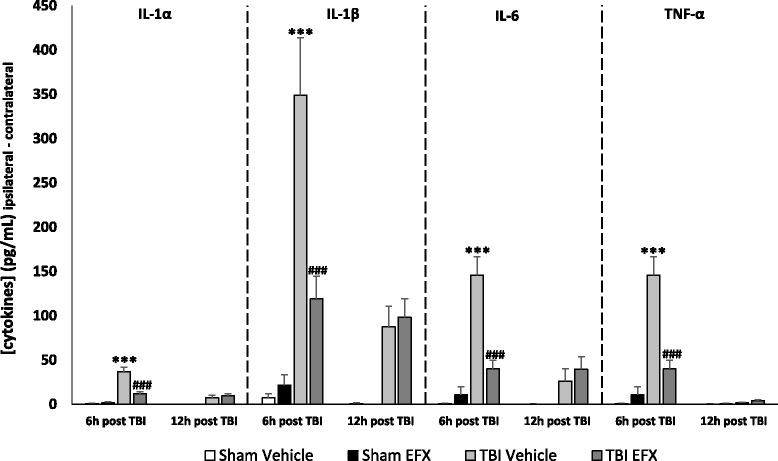
Cortical cytokine levels at 6 and 12 h post-CCI. The pro-inflammatory cytokines IL-1α, IL-1β, Il-6, and TNF-α were significantly elevated 6 h after CCI in the injury and margin regions compared to sham animals (***p < 0.001 for all groups). Statistical analyses showed no difference at 12-h time-point. Six hours after CCI, EFX treatment (50 mg/kg, i.p., 30 min after injury) significantly reduced IL-1α, IL-1β, IL-6, and TNF-α peaks (### p < 0.001 for all groups). ***p < 0.001: comparison between sham-vehicle and TBI-vehicle groups; ### p < 0.001: comparison between TBI-vehicle and TBI-EFX groups

Similar articles

Cited by

References

    1. Pop V, Badaut J. A neurovascular perspective for long-term changes after brain trauma. Transl Stroke Res. 2011;2:533–545. doi: 10.1007/s12975-011-0126-9. - DOI - PMC - PubMed
    1. Morganti-Kossmann MC, Rancan M, Stahel PF, Kossmann T. Inflammatory response in acute traumatic brain injury: a double-edged sword. Curr Opin Crit Care. 2002;8:101–105. doi: 10.1097/00075198-200204000-00002. - DOI - PubMed
    1. Chen SF, Hung TH, Chen CC, Lin KH, Huang YN, Tsai HC, et al. Lovastatin improves histological and functional outcomes and reduces inflammation after experimental traumatic brain injury. Life Sci. 2007;81:288–298. doi: 10.1016/j.lfs.2007.05.023. - DOI - PubMed
    1. Kumar A, Loane DJ. Neuroinflammation after traumatic brain injury: opportunities for therapeutic intervention. Brain Behav Immun. 2012;26:1191–1201. doi: 10.1016/j.bbi.2012.06.008. - DOI - PubMed
    1. Papadopoulos V, Lecanu L. Translocator protein (18 kDa) TSPO: an emerging therapeutic target in neurotrauma. Exp Neurol. 2009;219:53–57. doi: 10.1016/j.expneurol.2009.04.016. - DOI - PMC - PubMed

MeSH terms