. 2016 Nov;14(11):1054-1067.
doi: 10.1158/1541-7786.MCR-16-0167. Epub 2016 Aug 26.

Cooperative Dynamics of AR and ER Activity in Breast Cancer

Affiliations

Cooperative Dynamics of AR and ER Activity in Breast Cancer

Nicholas C D'Amato et al. Mol Cancer Res. 2016 Nov.

Abstract

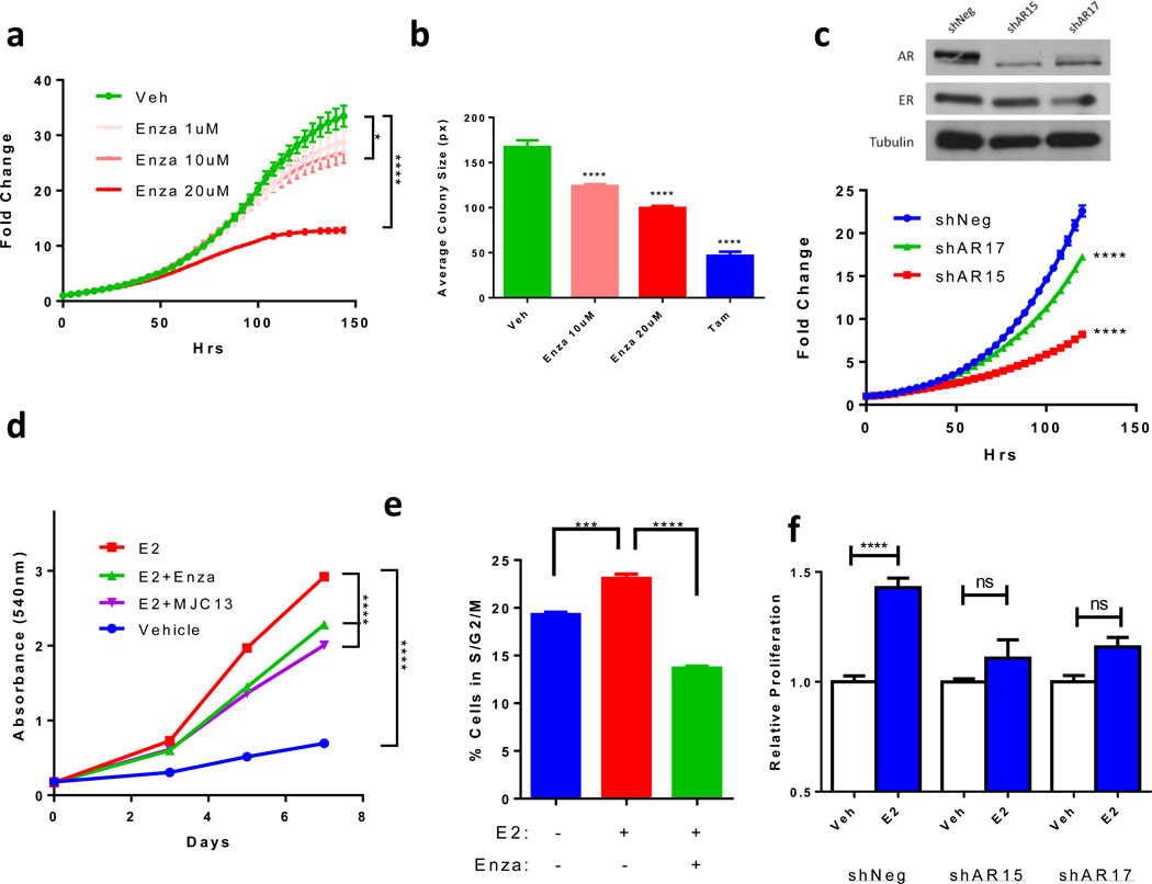
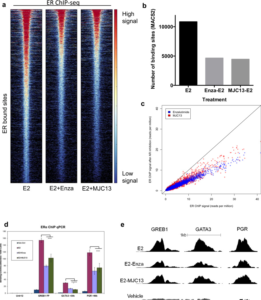
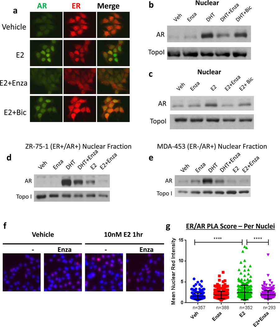
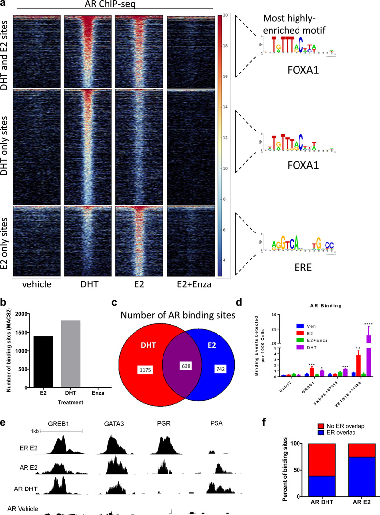
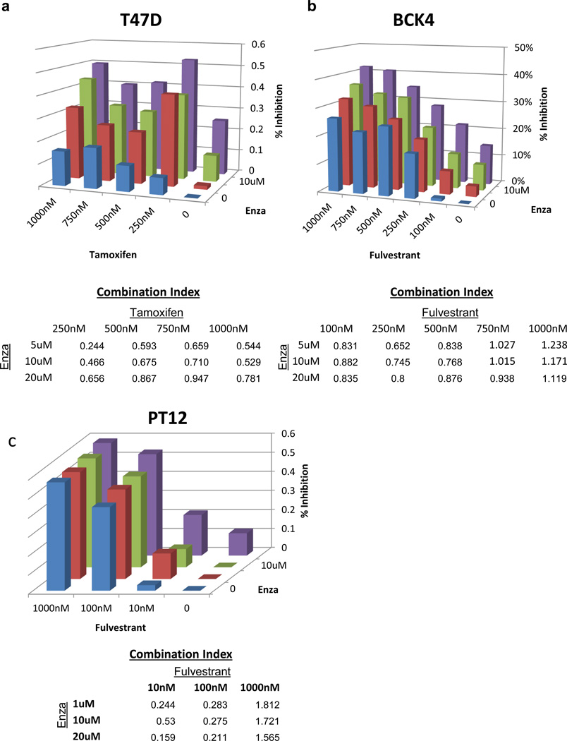
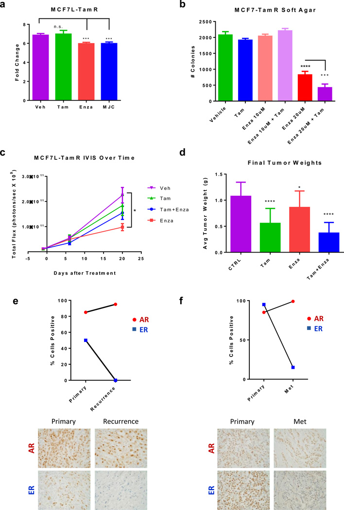
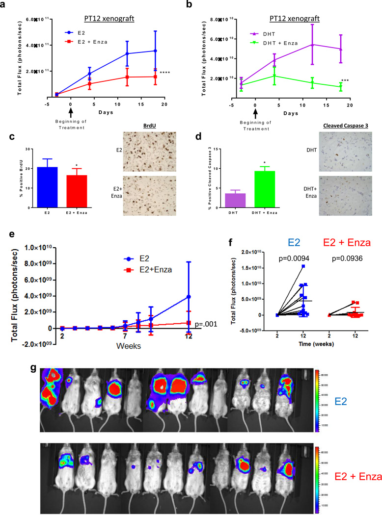
Androgen receptor (AR) is expressed in 90% of estrogen receptor alpha-positive (ER+) breast tumors, but its role in tumor growth and progression remains controversial. Use of two anti-androgens that inhibit AR nuclear localization, enzalutamide and MJC13, revealed that AR is required for maximum ER genomic binding. Here, a novel global examination of AR chromatin binding found that estradiol induced AR binding at unique sites compared with dihydrotestosterone (DHT). Estradiol-induced AR-binding sites were enriched for estrogen response elements and had significant overlap with ER-binding sites. Furthermore, AR inhibition reduced baseline and estradiol-mediated proliferation in multiple ER+/AR+ breast cancer cell lines, and synergized with tamoxifen and fulvestrant. In vivo, enzalutamide significantly reduced viability of tamoxifen-resistant MCF7 xenograft tumors and an ER+/AR+ patient-derived model. Enzalutamide also reduced metastatic burden following cardiac injection. Finally, in a comparison of ER+/AR+ primary tumors versus patient-matched local recurrences or distant metastases, AR expression was often maintained even when ER was reduced or absent. These data provide preclinical evidence that anti-androgens that inhibit AR nuclear localization affect both AR and ER, and are effective in combination with current breast cancer therapies. In addition, single-agent efficacy may be possible in tumors resistant to traditional endocrine therapy, as clinical specimens of recurrent disease demonstrate AR expression in tumors with absent or refractory ER.

Implications: This study suggests that AR plays a previously unrecognized role in supporting E2-mediated ER activity in ER+/AR+ breast cancer cells, and that enzalutamide may be an effective therapeutic in ER+/AR+ breast cancers. Mol Cancer Res; 14(11); 1054-67. ©2016 AACR.


Conflict of interest statement

Vernon T. Phan is a fulltime employee of Medivation, Inc.

Figures

Figure 1
Figure 1. AR inhibition decreases ER+/AR+ breast cancer growth
(a) Proliferation of MCF7 cells treated with increasing concentrations of Enza was monitored by IncuCyte. (b) MCF7 cells were grown in soft agar with Enza or tam and colony size was measured by ImageJ. (c) Immunoblotting for AR, ER, and Tubulin in MCF7 cells expressing a non-targeting (shNeg) or AR-targeting (shAR15 and shAR17) shRNA constructs (upper). Proliferation was monitored by IncuCyte (lower). (d) MCF7 cells were grown in media with CSS for 72 hrs then treated with vehicle (Veh), E2, or E2 + Enza or MJC13 and cell number was measured by crystal violet. (e) MCF7 cells were grown in media with CSS for 72hrs then treated with Veh, E2, or E2+Enza for 24hrs followed by cell cycle analysis. (f) MCF7 cells expressing shNeg, shAR15, or shAR17 were cultured in media with CSS for 72 hrs then treated with veh or E2 and growth was measured by crystal violet. Error bars represent standard error of the mean. * p<.05, **** p<.0001 by ANOVA with Dunnett’s Multiple Comparison Test.
Figure 2
Figure 2. AR inhibitors diminish ER genomic binding
ChIP-seq for ER in MCF7 cells grown in CSS for 3 days then treated with E2 +/− Enza or MJC-13. (a) Heat map of ER binding. The heat map is shown with a horizontal window of +/− 2kb. (b) The number of binding sites identified by MACS2, using vehicle treatment as the control. (c) The ER ChIP-seq signal at individual sites with E2 alone (x-axis) versus E2 + Enza (blue) or MJC13 (red) (y-axis). (D–E) ChIP-qPCR (d) and ChIP-seq read depth (e) at well-characterized ER binding sites. Error bars represent standard error of the mean. ** p<.01, *** p<.001, **** p<.0001 by ANOVA with Dunnett’s Multiple Comparison Test except.
Figure 3
Figure 3. AR and ER co-localize in the nucleus in response to E2
(a) MCF7 cells were grown in media with CSS for 72 hr then pre-treated with veh, 10 uM Enza, or 1 uM bicalutamide (bic). Following pre-treatment, cells were treated with veh or 10nM E2 +/− Enza or bic as shown for an additional 3 hr. Cells were then fixed and ICC was performed for AR (green) and ER (red). (b,c) MCF7 cells were grown in media with CSS for 72hrs then treated with the indicated treatment for 3 hrs, and nuclear extracts were immunoblotted for AR and TOPO1. (d) ER+/AR+ ZR-75-1 or (e) ER−/AR+ MDA-453 cells were grown in media with CSS for 72hrs, then pre-treated for 3 hr with Enza or vehicle control. Following pre-treatment, cells were treated with veh, 10nM DHT, or 10nM E2 +/− Enza as shown for 3 additional hrs. Nuclear extracts were then obtained and subjected to western blotting for AR and TopoI. (f) MCF7 cells were grown in media with CSS for 72hrs then treated with E2 +/− Enza for 1 h followed by fixation and PLA staining for AR and ER (red). Nuclei were stained with DAPI (blue). (g) Fluorescent intensity per nuclei was measured by CellProfiler. Error bars represent standard error of the mean. ****p <0.0001 by ANOVA with Dunnett’s Multiple Comparison Test.
Figure 4
Figure 4. E2 induces AR genome binding that overlaps with ER binding
ChIP-seq for AR in MCF7 cells grown in CSS for 3 days then treated with E2 for 1 h or DHT for 4 h. (a) Heat map of binding showing a horizontal window of +/− 2kb and enriched motifs from each category. (b) The number of binding sites identified by MACS2, using vehicle treatment as the control. (c) The number of AR binding sites that are unique to DHT (red), unique to E2 (blue), or shared (overlap) are shown. (d,e) ChIP-qPCR (d) and ChIP-seq read depth (e) results show AR binding at well-characterized ER binding sites following E2 treatment. (f) The percentage of AR binding sites in response to DHT (left) or E2 (right) that were also identified as ER binding sites (blue) is shown.
Figure 5
Figure 5. Enza synergizes with tam and fulvestrant in vitro
(a) T47D cells were grown in media with complete serum and Enza and/or tam, and cell number was monitored by IncuCyte. Percent inhibition was compared to vehicle after 5 days, and synergy was calculated using Calcusyn software. A Combination Index (CI) value < 1 is indicative of synergy (yellow). (b–c) BCK4 or PT12 cells were grown in phenol red-free media with CSS for 1 day then treated with E2 with Enza and/or fulvestrant and cell number was monitored by Incucyte.
Figure 6
Figure 6. Enza inhibits tamoxifen-resistant tumor growth in vitro and in vivo, and AR is expressed in recurrent breast cancers
(a) Growth of MCF7-TamR cells treated with vehicle, Tam, Enza, or MJC13 for 7 days. (b) MCF7-TamR cells were plated in soft agar and the number of colonies was counted after 14 days. (c–d) MCF7-TamR cells were implanted into the mammary glands of nude mice with estrogen pellets and were matched into groups to receive either control chow (CTRL), tamoxifen pellets (tam), enzalutamide-containing chow (enza), or both (tam+enza). (c) Tumor growth was measured over time by luminescence. (d) Final tumor weights of mice from each group (lower). (e) IHC for AR and ER in clinical samples of patient-matched primary tumor and recurrence 110 months later. (f) IHC for AR and ER in clinical samples of patient-matched primary tumor and metastasis 167 months later **(400×). * p<.05, *** p<.001, **** p<.0001 by ANOVA with Dunnett’s Multiple Comparison Test.
Figure 7
Figure 7. Enza decreases hormone driven growth of PT12 primary tumors and metastases
(a–d) 1×10⁶ GFP-luciferase expressing PT12 cells were injected orthotopically into the mammary fat pad of NOD-SCID-IL2Rgc−/− mice followed by implantation of either an E2 or DHT pellet. When tumors reached an average of 39 mm3, mice were randomized into the following groups: E2 with control chow (n=10) or enza chow (n=10), or DHT with control chow (n=5) or enza chow (n=5). Tumor viability was measured by IVIS for mice with E2 pellets (a) or DHT pellets (b). (c) BrdU staining of tumors from mice with E2 pellets. (d) Cleaved caspase staining of tumors from mice with DHT pellets. (e) PT12 GFP-luciferase cells were injected intracardially in NOD-SCID-IL2Rgc−/− mice. Metastatic burden of mice treated with E2 or E2+Enza was monitored using IVIS (photons/second) over 12 weeks (total signal supine+prone). (f) IVIS signal from mice in supine position at 2 weeks versus 12 weeks in E2 versus E2+Enza mice. (g) IVIS image of mice in the supine position in the E2 (n=11) or E2+Enza (n=12) groups after 12 weeks, red denotes high IVIS signal. Error bars represent SEM. *p<0.05, ***p<0.001, ****p <0.0001 by repeated measures mixed model approach for (a–b), ANOVA with Bonferroni’s Multiple Comparison Test for (c–d), and Wilcoxon rank sum test for (e).

References

    1. Collins LC, Cole KS, Marotti JD, Hu R, Schnitt SJ, Tamimi RM. Androgen receptor expression in breast cancer in relation to molecular phenotype: results from the Nurses' Health Study. Mod Pathol. 2011;24(7):924–931. - PMC - PubMed
    1. Peters AA, Buchanan G, Ricciardelli C, Bianco-Miotto T, Centenera MM, Harris JM, et al. Androgen receptor inhibits estrogen receptor-alpha activity and is prognostic in breast cancer. Cancer research. 2009;69(15):6131–6140. - PubMed
    1. Vera-Badillo FE, Templeton AJ, de Gouveia P, Diaz-Padilla I, Bedard PL, Al-Mubarak M, et al. Androgen receptor expression and outcomes in early breast cancer: a systematic review and meta-analysis. Journal of the National Cancer Institute. 2014;106(1):djt319. - PubMed
    1. Tsang JY, Ni YB, Chan SK, Shao MM, Law BK, Tan PH, et al. Androgen receptor expression shows distinctive significance in ER positive and negative breast cancers. Annals of surgical oncology. 2014;21(7):2218–2228. - PubMed
    1. Panet-Raymond V, Gottlieb B, Beitel LK, Pinsky L, Trifiro MA. Interactions between androgen and estrogen receptors and the effects on their transactivational properties. Molecular and Cellular Endocrinology. 2000;167:139–150. - PubMed

MeSH terms