. 2016 Aug 25;166(5):1198-1214.e24.
doi: 10.1016/j.cell.2016.07.027.

Two Distinct Types of E3 Ligases Work in Unison to Regulate Substrate Ubiquitylation

Affiliations

Two Distinct Types of E3 Ligases Work in Unison to Regulate Substrate Ubiquitylation

Daniel C Scott et al. Cell. .

Abstract

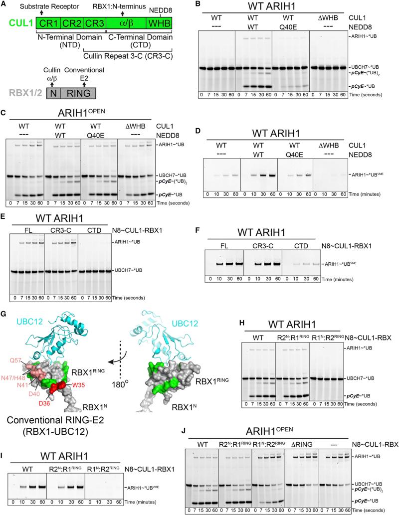
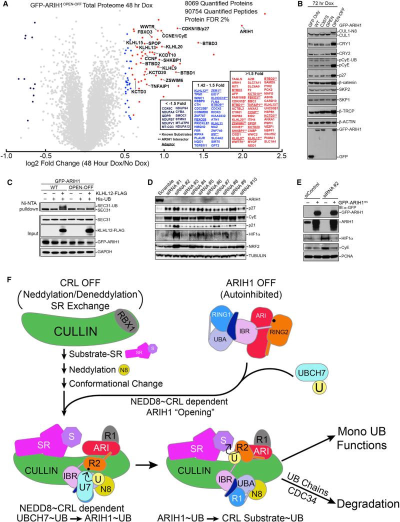
Hundreds of human cullin-RING E3 ligases (CRLs) modify thousands of proteins with ubiquitin (UB) to achieve vast regulation. Current dogma posits that CRLs first catalyze UB transfer from an E2 to their client substrates and subsequent polyubiquitylation from various linkage-specific E2s. We report an alternative E3-E3 tagging cascade: many cellular NEDD8-modified CRLs associate with a mechanistically distinct thioester-forming RBR-type E3, ARIH1, and rely on ARIH1 to directly add the first UB and, in some cases, multiple additional individual monoubiquitin modifications onto CRL client substrates. Our data define ARIH1 as a component of the human CRL system, demonstrate that ARIH1 can efficiently and specifically mediate monoubiquitylation of several CRL substrates, and establish principles for how two distinctive E3s can reciprocally control each other for simultaneous and joint regulation of substrate ubiquitylation. These studies have broad implications for CRL-dependent proteostasis and mechanisms of E3-mediated UB ligation.


Figures

Figure 1
Figure 1. ARIH1 Broadly Interacts with Components of Assembled, Neddylated CRLs
(A) Left to right: immunoblot of low-level induced expression of FLAG-HA-ARIH1 or catalytic C375S mutant in 293 FLP-IN cells; high-confidence interacting proteins (HCIPs) for WT ARIH1 and ARIH1C357S, ± neddylation inhibitor MLN4924, identified by CompPASS (Sowa et al., 2009). ARIH1 predominantly interacts with CRL subunits colored by CRL1, blue; CRL2, yellow; CRL3, purple. (B) TMT method to identify MLN4924-sensitive/neddylation-dependent ARIH1 HCIPs. (C) ARIH1C357S HCIPs decreased ≥2-fold by neddylation inhibitor MLN4924 colored by CRL: CRL1, blue; CRL2, yellow; CRL3, violet. CRL3KLHL12 and substrate SEC13-SEC31A are highlighted. Error, ±SEM. (D) Log2 fold change of ARIH1C357S interactors from the schematic in (C). Error bars, ±SE.
Figure 2
Figure 2. ARIH1 Rapidly, Efficiently, and Specifically Mediates UB Ligation to CRL Substrates
(A) Pulse-chase reaction scheme. Pulse: thioester-linked UBCH7~*UB is generated and E1 reaction is quenched. Chase: ARIH1 is added; path of fluorescent UB* via ARIH1 is followed in presence or absence of neddylated or non-neddylated CUL-RBX1, SR, and CRL substrate indicated in bold. (B) Fluorescent scans of gels of assay in (A), showing neddylated CRL3KLHL12 relieves ARIH1 autoinhibition, promotes autoubiquitylation in absence of CRL substrate, and UB ligation to CRL substrate SEC31A depending on neddylation, the SR KLHL12, and ARIH1 catalytic Cys357. (C) Inferred pathway from (B) and generalization: ARIH1 mediates UB ligation to CRL client substrates depending on CUL neddylation, the substrate receptor, and ARIH1's catalytic Cys. (D) Fluorescent scans of gels of assay in (A), showing neddylated CRL1FBW7ΔD relieves ARIH1 autoinhibition, promotes autoubiquitylation in absence of CRL substrate, and UB ligation to CRL peptide substrate pCyE depending on neddylation (N8~CUL1), SR, and ARIH1 catalytic Cys. (E) Assays showing UBCH7-ARIH1-mediated UB ligation to pCyE as a function CRL1FBW7ΔD concentration for kinetic analyses. (F) Michaelis-Menten analysis of data from (E) to calculate kinetic parameters for ARIH1-mediated UB ligation to pCyE via neddylated CRL1FBW7ΔD.
Figure 3
Figure 3. ARIH1-CRL Mechanism Efficiently Mediates monoUB Ligation to CUL3 and CUL1 Substrates
(A) Pulse-chase assays starting with indicated E2~*UB or~*UB K0 (cannot form Lys-linked polyUB chains) transfer to CRL3KLHL12 substrate SEC31A via UBCH7 by ARIH1-CRL pathway or with E2s UBCH5B or CDC34B by canonical CRL RING mechanism. Right, quantification of total *UB or *UB K0 transfer to SEC31A. (B) As (A), but for CRL1FBW7ΔD and client substrate pCyE. (C) As (A), but for CRL1FBXL3 and client substrate CRY1. (D) Scheme of two-color pulse-chase assay tracking UB (WT or K0) transferred from either UBCH7 or CDC34B in the same tube to NEDD8~CRL1FBXL3 client substrate CRY1. In pulse, UBCH7 was loaded with “green” *UB and CDC34 with “red” *UB or *UB K0, and E1 reaction was quenched. Distinguishing green-, yellow-, orange-, and red-colored products in chase reaction allows distinguishing individual E2 reactions and sequential *UB priming by ARIH1/UBCH7 and chain extension by CDC34B. (E) Reactions from (D) simultaneously monitoring UBCH7 and CDC34-mediated *UB transfer to CRL1FBXL3 client CRY1 indicates priming by UBCH7-ARIH1 and extending by CDC34.
Figure 4
Figure 4. Broad Mutational Survey Reveals Numerous ARIH1 Surfaces Contribute to CRL Substrate Ubiquitylation
(A) ARIH1 domains. (B) Sites of Ala mutations as spheres on structure of autoinhibited ARIH1 (Duda et al., 2013), colored by effect on *UB transfer from UBCH7 via ARIH1/neddylated CRL1FBW7ΔD to pCyE (in Figure S4): gray, normal; green, hyperactive; blue, ligation defect/accumulation of normally transient thioester-bonded ARIH1~UB intermediate; raspberry, marginal defect; red, strong defect. (C) Most defective (red) and ligation-impaired (blue) ARIH1 Ala scan mutants in pulse-chase assays monitoring entire pathway: *UB transfer from UBCH7 via the indicated ARIH1 mutants and neddylated CRL1FBW7ΔD to pCyE. Ligation-impaired mutants are scored by appearance and dissipation of ARIH1~UB thioester intermediate.
Figure 5
Figure 5. Activity Profiling of ARIH1 Mutants: “Signatures” Identifying Functions in UB Ligation to Neddylated CRL Substrates
(A) Hierarchical assay scheme to profile selected ARIH1 mutants in neddylated CRL pathway. The sum of results from all assays form a signature, or “fingerprint” identifying normal function(s) of a mutated surface. Color coding of functions is maintained in structures and signatures in (B)–(I), except signatures VII and VIII that signify dual functions (orange/purple and orange/red). Details are in Figure S5. (B) Close up showing ARIH1 catalytic Cys that is normally sequestered by autoinhibitory intramolecular RING2-Ariadne (ARI) domain interface (4KBL.PDB). Hyperactive mutant sites are spheres: magenta, relieve autoinhibition even without neddylated CRL, thus termed “OPEN” due to mutationally opening the RING2-ARI interface; cyan, facilitate “opening” by neddylated CRL, monitored by reactivity with *UB-VME. OPEN mutant B was combined with other mutations for assays 3, 4, and 5 in subsequent profiles. (C) Activity profile signatures of hyperactive mutants, normalized to WT ARIH1, defined by assays 1–3. (D) Model of ARIH1 RING2~UB intermediate based on 4KBL.PDB and 4LJO.PDB. Sites of activity profile signatures indicating roles in catalytic UB binding or intrinsic ligation activity are in blue and purple spheres, respectively. (E) Activity profile signatures for mutants in (D). (F) ARIH1's RING1 and UBA domains modeled with docked E2 or UBL (NEDD8/UB), respectively (4KBL.PDB, 5EDV.PDB, 4UN2.PDB). Sites of mutants with activity profile signatures indicating roles in UB transfer from E2 (UBCH7) to ARIH1 and neddylated CRL-dependent UB transfer from UBCH7 to ARIH1 are in gray and green spheres, respectively. (G) Activity profile signatures for mutants in (F). (H) ARIH1 structure as in (B), rotated ≈60° about x, highlighting residues in RING2 and ARI domains with dual roles in two colors: neddylated CUL-dependent relief of ARIH1 autoinhibition, orange; role in intrinsic RING2-mediated UB ligation to a Lys nucleophile, purple; role in specifically targeting CRL substrate, red. (I) Activity profile signatures for mutants in (H).
Figure 6
Figure 6. Distinctive and Specialized Roles of Neddylation, CUL1, and RBX1 in Employing ARIH1 to Target a CRL Substrate
(A) CUL1 and RBX domains (Zheng et al., 2002), denoting previously known functional interactions. (B) Pulse-chase assay as in Figure 2A testing role of neddylation in UBCH7-ARIH1-mediated *UB transfer to pCyE with indicated versions of CRL1FBW7ΔD (± neddylation, with NEDD8 Q40E mutant that does not support CUL1-RBX1 conformational change, or with CUL1 ΔWHB lacking domain with neddylation site). (C) Assay as in (B) except with ARIH1OPEN mutant that is not autoinhibited. ARIH1OPEN overcomes defects from lack of neddylation, indicating role of CRL neddylation in relieving ARIH1 autoinhibition. (D) Roles of neddylation in promoting ARIH1 “opening” as probed reactivity with *UB-VME, showing defect caused by Q40E and that neddylation is not mimicked by deleting CUL1's WHB domain. (E) Role of CUL1 N-terminal domain, specifically the CR3 subdomain, in relieving ARIH1 autoinhibition, tested by ARIH1 autoubiquitylation in presence of indicated versions of neddylated CUL1-RBX1 (no SR, no substrate). (F) Role of CUL1 N-terminal domain, specifically the CR3 subdomain, in stimulating ARIH1 “opening,” probed by ability of indicated versions of neddylated CUL1-RBX1 to stimulate ARIH1 reactivity with *UB-VME. (G) Effects of RBX1 mutants on ARIH1-mediated ubiquitylation of neddylated CRL substrate, mapped on conventional RING E3-E2 (RBX1-UBC12) structure (Scott et al., 2014). Green, normal; raspberry, marginal defect; red, strong defect (data in Figure S6A). (H) Role of RBX1 RING on ARIH1-neddylated CRL pathway tested by RBX1/RBX2 domain swaps, assayed as in (B). (I) Role of RBX1 RING in neddylated CUL1-RBX1-dependent “opening” of ARIH1, assayed as in (D). (J) Second, the distinctive role of RBX1 RING (directing UB transfer from ARIH1 to neddylated CRL substrate) uncovered by use of ARIH1OPEN mutant. In the absence of autoinhibition, this assay directly tests CRL substrate (pCyE) targeting by the indicated RING mutations in neddylated CRL1FBW7ΔD. Right-most lanes, control confirming ARIH1OPEN autoubiquitylation in absence of neddylated CRL.
Figure 7
Figure 7. Widespread Effects of Dominant-Negative ARIH1OPEN-OFF or ARIH1 Knockdown on CRL Pathways and Model for Extreme Amalgamation of ARIH1 and Neddylated CRLs in Mediating Regulation
(A) TMT-based quantification of the total proteome following expression of GFP-ARIH1OPEN-OFF for 48 hr. Whole-cell lysates were prepared in triplicate for each condition and pooled following TMT labeling. ≥1.5-fold enrichment in GFP-ARIH1OPEN-OFF expressing cells, red, 1.42- to 1.5-fold increase, blue. *Previously reported CRL substrates. **ARIH1 interactors. (B) Immunoblots of indicated CRL1 substrates following expression of GFP-ARIH1OPEN-OFF for 72 hr. (C) Overexpression of GFP-ARIHOPEN-OFF inhibits cellular ubiquitylation of SEC31A. (D) Depletion of ARIH1 in 293T cells with ten different siRNAs for 72 hr increases levels of CRL substrates detected by indicated immunoblots. (E) Expressing siRNA-resistant GFP-ARIH1 reverses increase in indicated CRL substrate levels after ARIH1 knockdown in 293T cells. (F) Model for ARIH1 and neddylated CRL acting in unison for joint E3-E3-mediated substrate ubiquitylation. In the absence of a substrate, CRL is OFF (unneddylated, unassembled, and inhibited) and ARIH1 is OFF (autoinhibited). Substrate binding to SR stimulates the pathway, promoting CRL assembly, neddylation, and conformational change, which, in turn, enables NEDD8, the cullin, and RBX1 to activate ARIH1 via ARIH1's UBA, RING2, and Ariadne (ARI) domains. This “opens” the ARIH1 structure and allows UB transfer from UBCH7 to ARIH1's RING2 catalytic Cys. Subsequently, specific surfaces from the CRL's RBX1 RING domain and from ARIH1's Ariadne domain promote monoUB transfer from ARIH1's RING2 catalytic Cys to the CRL's substrate receptor (SR)-bound substrate. The monoubiquitylated substrate is primed for altered function, further multimonoubiquitylation via repeated cycles through the ARIH1-CRL path, polyubiquitylation via a canonical RING-E2 mechanism directing proteasomal degradation, or other fates.

Comment in

  • Tag Team Ubiquitin Ligases.
    Kleiger G, Deshaies R. Kleiger G, et al. Cell. 2016 Aug 25;166(5):1080-1081. doi: 10.1016/j.cell.2016.08.014. Cell. 2016. PMID: 27565338

Similar articles

Cited by

References

    1. Bennett EJ, Rush J, Gygi SP, Harper JW. Dynamics of cullin-RING ubiquitin ligase network revealed by systematic quantitative proteomics. Cell. 2010;143:951–965. - PMC - PubMed
    1. Berndsen CE, Wolberger C. New insights into ubiquitin E3 ligase mechanism. Nat. Struct. Mol. Biol. 2014;21:301–307. - PubMed
    1. Busino L, Bassermann F, Maiolica A, Lee C, Nolan PM, Godinho SI, Draetta GF, Pagano M. SCFFbxl3 controls the oscillation of the circadian clock by directing the degradation of cryptochrome proteins. Science. 2007;316:900–904. - PubMed
    1. Ceccarelli DF, Tang X, Pelletier B, Orlicky S, Xie W, Plantevin V, Neculai D, Chou YC, Ogunjimi A, Al-Hakim A, et al. An allosteric inhibitor of the human Cdc34 ubiquitin-conjugating enzyme. Cell. 2011;145:1075–1087. - PubMed
    1. Deshaies RJ, Joazeiro CA. RING domain E3 ubiquitin ligases. Annu. Rev. Biochem. 2009;78:399–434. - PubMed

Publication types