Diagnostic value of qualitative and strain ratio elastography in the differential diagnosis of non-palpable testicular lesions
- PMID: 27565451
- PMCID: PMC5108442
- DOI: 10.1111/andr.12260
Diagnostic value of qualitative and strain ratio elastography in the differential diagnosis of non-palpable testicular lesions
Abstract
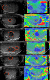
The purpose of this study was to evaluate prospectively the accuracy of qualitative and strain ratio elastography (SE) in the differential diagnosis of non-palpable testicular lesions. The local review board approved the protocol and all patients gave their consent. One hundred and six patients with non-palpable testicular lesions were consecutively enrolled. Baseline ultrasonography (US) and SE were correlated with clinical and histological features and ROC curves developed for diagnostic accuracy. The non-palpable lesions were all ≤1.5 cm; 37/106 (34.9%) were malignant, 38 (35.9%) were benign, and 31 (29.2%) were non-neoplastic. Independent risk factors for malignancy were as follows: size (OR 17.788; p = 0.002), microlithiasis (OR 17.673, p < 0.001), intralesional vascularization (OR 9.207, p = 0.006), and hypoechogenicity (OR, 11.509, p = 0.036). Baseline US had 89.2% sensitivity (95% CI 74.6-97.0) and 85.5% specificity (95% CI 75.0-92.8) in identifying malignancies, and 94.6% sensitivity (95% CI 86.9-98.5) and 87.1% specificity (95% CI 70.2-96.4) in discriminating neoplasms from non-neoplastic lesions. An elasticity score (ES) of 3 out of 3 (ES3, maximum hardness) was recorded in 30/37 (81.1%) malignant lesions (p < 0.001). An intermediate score of 2 (ES2) was recorded in 19/38 (36.8%) benign neoplastic lesions and in 22/31 (71%) non-neoplastic lesions (p = 0.005 and p = 0.001 vs. malignancies). None of the non-neoplastic lesions scored ES3. Logistic regression analysis revealed a significant association between ES3 and malignancy (χ2 = 42.212, p < 0.001). ES1 and ES2 were predictors of benignity (p < 0.01). Overall, SE was 81.8% sensitive (95% CI 64.8-92.0) and 79.1% specific (95% CI 68.3-88.4) in identifying malignancies, and 58.6% sensitive (95% CI 46.7-69.9) and 100% specific (95% CI 88.8-100) in discriminating non-neoplastic lesions. Strain ratio measurement did not improve the accuracy of qualitative elastography. Strain ratio measurement offers no improvement over elastographic qualitative assessment of testicular lesions; testicular SE may support conventional US in identifying non-neoplastic lesions when findings are controversial, but its added value in clinical practice remains to be proven.
Keywords: Leydig cell tumor; male infertility; scrotal ultrasound; seminoma; strain elastography; testicular cancer.
© 2016 The Authors. Andrology published by John Wiley & Sons Ltd on behalf of American Society of Andrology and European Academy of Andrology.
Figures


References
-
- Aigner F, De Zordo T, Pallwein‐Prettner L, Junker D, Schafer G, Pichler R, Leonhartsberger N, Pinggera G, Dogra VS & Frauscher F. (2012) Real‐time sonoelastography for the evaluation of testicular lesions. Radiology 263, 584–589. - PubMed
-
- American Institute of Ultrasound in Medicine , American College of Radiology , Society of Radiologists in Ultrasound . (2011) AIUM practice guideline for the performance of scrotal ultrasound examinations. J Ultrasound Med 30, 151–155. - PubMed
-
- Appelbaum L, Gaitini D & Dogra VS. (2013) Scrotal ultrasound in adults. Semin Ultrasound CT MR 34, 257–273. - PubMed
-
- Bhatt S, Rubens DJ & Dogra VS. (2006) Sonography of benign intrascrotal lesions. Ultrasound Q 22, 121–136. - PubMed
-
- Brock M, von Bodman C, Palisaar RJ, Loppenberg B, Sommerer F, Deix T, Noldus J & Eggert T. (2012) The impact of real‐time elastography guiding a systematic prostate biopsy to improve cancer detection rate: a prospective study of 353 patients. J Urol 187, 2039–2043. - PubMed
Publication types
MeSH terms
LinkOut - more resources
Full Text Sources
Other Literature Sources
Medical
Molecular Biology Databases
Miscellaneous

