. 2016 Aug 26;16(1):195.
doi: 10.1186/s12866-016-0814-4.

Synergism and the mechanism of action of the combination of α-mangostin isolated from Garcinia mangostana L. and oxacillin against an oxacillin-resistant Staphylococcus saprophyticus

Affiliations

Synergism and the mechanism of action of the combination of α-mangostin isolated from Garcinia mangostana L. and oxacillin against an oxacillin-resistant Staphylococcus saprophyticus

Sineewan Phitaktim et al. BMC Microbiol. .

Abstract

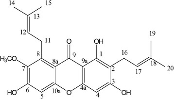
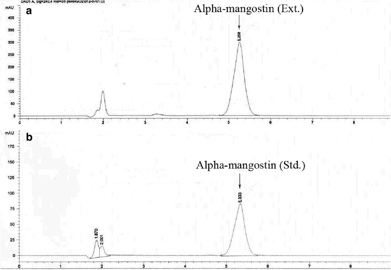
Background: Globally, staphylococci have developed resistance to many antibiotics. New approaches to chemotherapy are needed and one such approach could be to use plant derived actives with conventional antibiotics in a synergestic way. The purpose of this study was to isolate α-mangostin from the mangosteen (Garcinia mangostana L.; GML) and investigate antibacterial activity and mechanisms of action when used singly and when combined with oxacillin against oxacillin-resistant Staphylococcus saprophyticus (ORSS) strains. The isolated α-mangostin was confirmed by HPLC chromatogram and NMR spectroscopy. The minimum inhibitory concentration (MIC), checkerboard and killing curve were determined. The modes of action of these compounds were also investigated by enzyme assay, transmission electron microscopy (TEM), confocal microscopic images, and cytoplasmic membrane (CM) permeabilization studies.

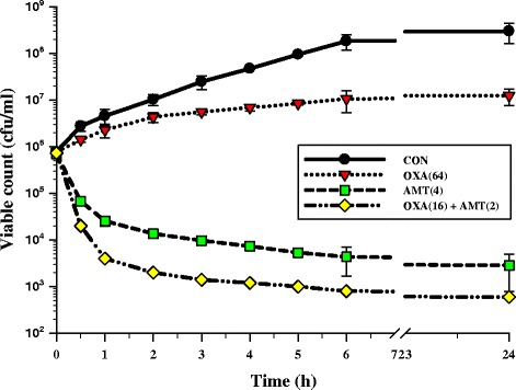
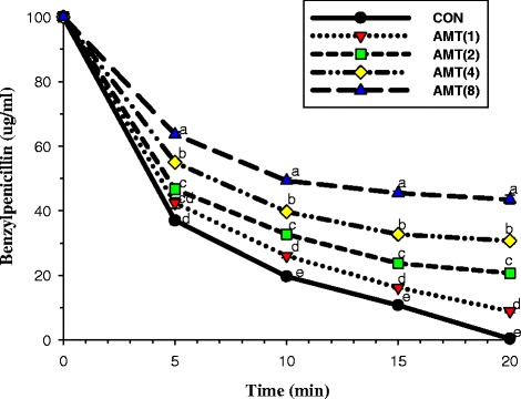
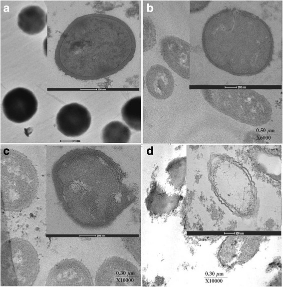
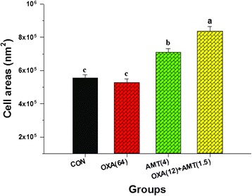
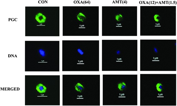
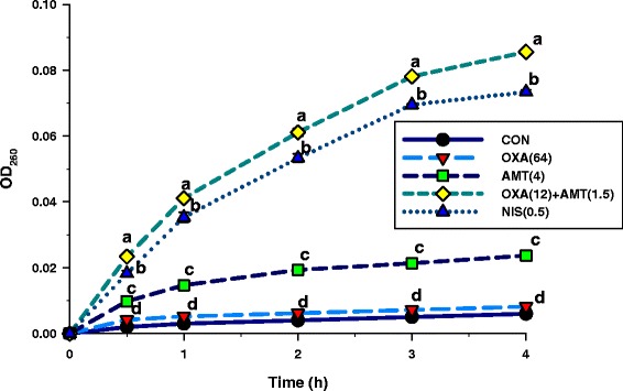
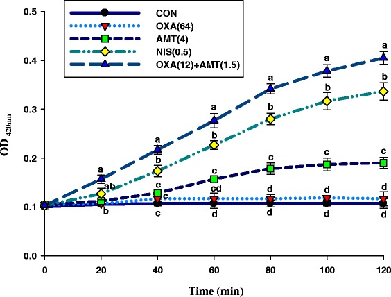
Results: The MICs of isolated α-mangostin and oxacillin against these strains were 8 and 128 μg/ml, respectively. Checkerboard assays showed the synergistic activity of isolated α-mangostin (2 μg/ml) plus oxacillin (16 μg/ml) at a fractional inhibitory concentration index (FICI) of 0.37. The kill curve assay confirmed that the viability of oxacillin-resistant Staphylococcus saprophyticus DMST 27055 (ORSS-27055) was dramatically reduced after exposure to isolated α-mangostin (2 μg/ml) plus oxacillin (16 μg/ml). Enzyme assays demonstrated that isolated α-mangostin had an inhibitory activity against β-lactamase in a dose-dependent manner. TEM results clearly showed that these ORSS-27055 cells treated with this combination caused peptidoglycan and cytoplasmic membrane damage, irregular cell shapes and average cell areas were significantly larger than the control. Clearly, confocal microscopic images confirmed that this combination caused considerable peptidoglycan damage and DNA leakage. In addition, the CM permeability of ORSS-27055 was also increased by this combination of actives.

Conclusions: These findings provide evidence that isolated α-mangostin alone has not only some activity but also shows the synergistic activity with oxacillin against ORSS-27055. The chromone and isoprenyl structures could play a significant role in its action. This synergistic activity may involve three mechanisms of action. Firstly, potential effects of cytoplasmic membrane disruption and increases permeability. Secondly, inhibit β-lactamase activity. Finally, also damage to the peptidoglycan structure. We proposes the potential to develop a novel adjunct phytopharmaceutical to oxacillin for the treatment of ORSS. Future studies require clinical trials to establish if the synergy reported can be translated to animals and humans.

Keywords: Garcinia mangostana; Mechanism of action; Oxacillin; Oxacillin-resistant S. saprophyticus; Synergistic activity; α-mangostin.


Figures

Fig. 1
Fig. 1
Structure of α-mangostin
Fig. 2
Fig. 2
HPLC chromatograms of α-mangostin: a, Alpha-mangostin (Ext.) = α-mangostin extract from G. mangostana; b, Alpha-mangostin (Std.) = α-mangostin standard
Fig. 3
Fig. 3
The effect of oxacillin, α-mangostin alone or in combination on the viable counts of ORSS-27055. CON = control (drugs free); OXA(64) = 64 μg/ml oxacillin; AMT(4) = 4 μg/ml α-mangostin; OXA(16) + AMT(2) = 16 μg/ml oxacillin plus 2 μg/ml α-mangostin. The values plotted are the means of 4 observations, and the vertical bars indicate the standard errors of the means
Fig. 4
Fig. 4
The inhibitory activity of α-mangostin against β-lactamase in hydrolysing benzylpenicillin. β-lactamase used from E. cloacae; CON = control (no testing agent); AMT(1) = 1 μg/ml α-mangostin. The graph shows the remaining benzylpenicillin at the same time. Means sharing the same superscript are not significantly different from each other (Scheffe’s test, p < 0.01)
Fig. 5
Fig. 5
Ultrathin sections of log phase oxacillin-resistant S. saprophyticus DMST 27055 grown in CAMHB containing: a, control (drug-free); b, oxacillin at 64 μg/ml; c, α-mangostin at 4 μg/ml; d, oxacillin at 12 μg/ml plus α-mangostin at 1.5 μg/ml; (Magnification; a, 4000×, bar = 1 μm; b, 6000×, bar = 0.5 μm; c, 10,000×, bar = 0.5 μm; d, 10,000×, bar = 0.3 μm; Inset magnification; a, c, 38,000×; b, 29,000×; d, 10,000×; bar; a, b, c, 200 nm; d, 500 nm)
Fig. 6
Fig. 6
The effect of either oxacillin or α-mangostin on average cross-section of ORSS-27055 cell areas. CON = control (drugs free); OXA(64) = 64 μg/ml oxacillin; AMT(4) = 4 μg/ml α-mangostin; OXA(12) + AMT(1.5) = 12 μg/ml oxacillin plus 1.5 μg/ml α-mangostin. The mean ± SEM for three replicates are illustrated. Means sharing the same superscript are not significantly different from each other (Scheffe’s test, p < 0.01)
Fig. 7
Fig. 7
Schematic representation of the results of immunofluorescence and a confocal laser scanning microscope; Samples of oxacillin-resistant S. saprophyticus DMST 27055 after treatment for 4 h with oxacillin, α-mangostin either alone or in combination. CON = control (drugs free); OXA(64) = 64 μg/ml oxacillin; AMT(4) = 4 μg/ml α-mangostin; OXA(12) + AMT(1.5) = 12 μg/ml oxacillin plus 1.5 μg/ml α-mangostin. The cells were stained for DNA with DAPI (blue, DNA) and labelled for peptidoglycan (green, PGC) using respective antibodies. DNA in all groups was localized in the central of the cell and surrounded by a peptidoglycan layer (MERGED). Scale bar = 1 μm
Fig. 8
Fig. 8
Effects of 260 nm absorbing material (DNA, RNA, and metabolites) in the ORSS-27055 supernatants. These cells were treated with oxacillin, α-mangostin either alone or in combination. CON = control (drugs free); OXA(64) = 64 μg/ml oxacillin; AMT(4) = 4 μg/ml α-mangostin; OXA(12) + AMT(1.5) = 12 μg/ml oxacillin plus 1.5 μg/ml α-mangostin; NIS(0.5) = 0.50 μg/ml nisin. Nisin at 0.50 μg/ml was used as a positive control, and untreated cells were used as a negative control. The mean ± SEM for three replicates are illustrated. Means sharing the same superscript at the same time are not significantly different (Scheffe’s test, p < 0.01)
Fig. 9
Fig. 9
Time-dependency of oxacillin plus α-mangostin-induced permeabilisation of the cytoplasmic membrane of ORSS-27055. The kinetics of α-mangostin-mediated ONPG passage across the cytoplasmic membrane are monitored as a change in the optical density (OD). The concentration of substrates was 100 μg/ml ONPG. CON = control (drugs free); OXA(64) = 64 μg/ml oxacillin; AMT(4) = 4 μg/ml α-mangostin; OXA(12) + AMT(1.5) = 12 μg/ml oxacillin plus 1.5 μg/ml α-mangostin; NIS(0.5) = 0.50 μg/ml nisin. Nisin at 0.50 μg/ml was used as a positive control, and untreated cells were used as a negative control. Each treatment performed two times in triplicate. The graph shows OD420nm of each treatment at the same time. Means sharing the same superscript at the same time are not significantly different from each other (Scheffe’s test, p < 0.01)
Fig. 10
Fig. 10
The effect of oxacillin, α-mangostin either alone or in combination on 3T3-L1 preadipocytes. 3T3-L1 preadipocytes were treated with: a. oxacillin (0-2048 μg/ml); b. α-mangostin (0-128 μg/ml); and c. oxacillin plus α-mangostin (8 + 1 to 512 + 64 μg/ml), for 24 h. Results are expressed as percentages of cell viability as compared with untreated controls (n = 8). The mean ± SEM for three replicates are illustrated. Means sharing the same superscript are not significantly different from each other (Scheffe’s test, p < 0.01)

References

    1. Huttner A, Harbarth S, Carlet J, Cosgrove S, Goossens H, Holmes A, et al. Antimicrobial resistance: a global view from the 2013 World Healthcare-Associated Infections Forum. Antimicrob Resist Infect Control. 2013;2:31. doi: 10.1186/2047-2994-2-31. - DOI - PMC - PubMed
    1. Rukachaisirikul V, Arunpanichlert J, Sukpondma Y, Phongpaichit S, Sakayaroj J. Metabolites from the endophytic fungi Botryosphaeria rhodina PSU-M35 and PSU-M114. Tetrahedron. 2009;65:10590–5. doi: 10.1016/j.tet.2009.10.084. - DOI
    1. Var SK, Hadi R, Khardori NM. Evaluation of regional antibiograms to monitor antimicrobial resistance in Hampton Roads, Virginia. Ann Clin Microbiol Antimicrob. 2015;14:22. doi: 10.1186/s12941-015-0080-6. - DOI - PMC - PubMed
    1. Widerstrom M, Wistrom J, Ferry S, Karlsson C, Monsen T. Molecular epidemiology of Staphylococcus saprophyticus isolated from women with uncomplicated community-acquired urinary tract infection. J Clin Microbiol. 2007;45:1561–4. doi: 10.1128/JCM.02071-06. - DOI - PMC - PubMed
    1. Higashide M, Kuroda M, Omura CT, Kumano M, Ohkawa S, Ichimura S, et al. Methicillin-resistant Staphylococcus saprophyticus isolates carrying staphylococcal cassette chromosome mec have emerged in urogenital tract infections. Antimicrob Agents Chemother. 2008;52:2061–8. doi: 10.1128/AAC.01150-07. - DOI - PMC - PubMed

Publication types

MeSH terms