. 2016 Oct 14;291(42):22315-22326.
doi: 10.1074/jbc.M116.744532. Epub 2016 Aug 26.

Post-translational Acetylation of MbtA Modulates Mycobacterial Siderophore Biosynthesis

Affiliations

Post-translational Acetylation of MbtA Modulates Mycobacterial Siderophore Biosynthesis

Olivia Vergnolle et al. J Biol Chem. .

Abstract

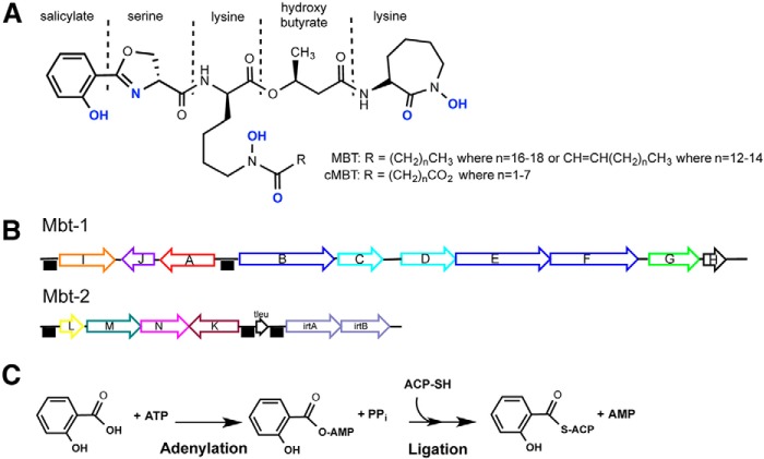
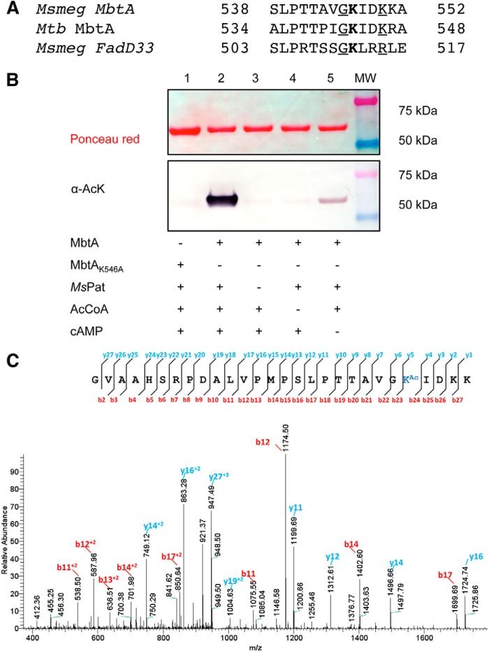
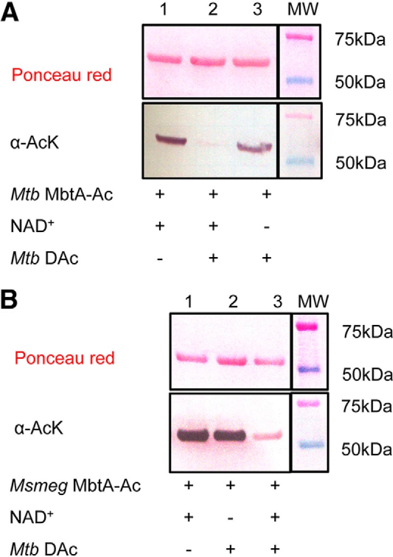
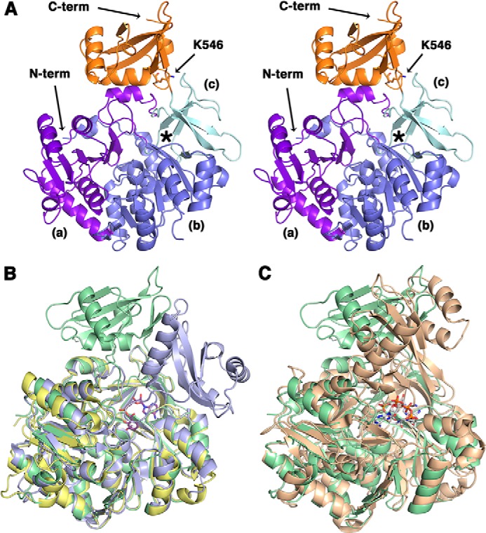
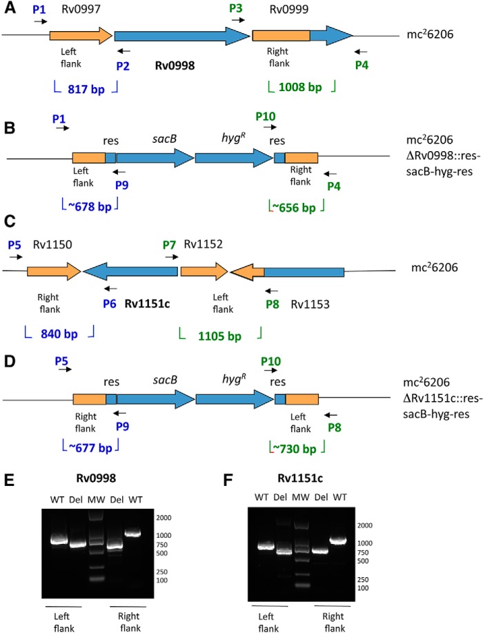
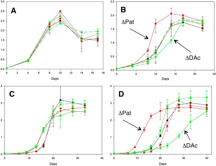
Iron is an essential element for life, but its soluble form is scarce in the environment and is rarer in the human body. Mtb (Mycobacterium tuberculosis) produces two aryl-capped siderophores, mycobactin (MBT) and carboxymycobactin (cMBT), to chelate intracellular iron. The adenylating enzyme MbtA catalyzes the first step of mycobactin biosynthesis in two half-reactions: activation of the salicylic acid as an acyl-adenylate and ligation onto the acyl carrier protein (ACP) domain of MbtB to form covalently salicylated MbtB-ACP. We report the first apo-MbtA structure from Mycobacterium smegmatis at 2.3 Å. We demonstrate here that MbtA activity can be reversibly, post-translationally regulated by acetylation. Indeed the mycobacterial Pat (protein lysine acetyltransferase), Rv0998, specifically acetylates MbtA on lysine 546, in a cAMP-dependent manner, leading to enzyme inhibition. MbtA acetylation can be reversed by the NAD+-dependent DAc (deacetyltransferase), Rv1151c. Deletion of Pat and DAc genes in Mtb revealed distinct phenotypes for strains lacking one or the other gene at low pH and limiting iron conditions. This study establishes a direct connection between the reversible acetylation system Pat/DAc and the ability of Mtb to adapt in limited iron conditions, which is critical for mycobacterial infection.

Keywords: MbtA; Mycobacterium tuberculosis; acetylation; enzyme structure; iron; mycobactin; post-translational modification (PTM).


Figures

FIGURE 1.
FIGURE 1.
Salicyl-capped mycobactin siderophore structure, genetic loci and first biosynthesis reaction. A, lipophilic MBT and hydrophilic cMBT share a common core structure but differ in the length of the alkyl substitution (R group). Atoms in blue are involved in the hexadentate ferric iron coordination. B, mbt-1 gene cluster produces mycobactin core, whereas mbt-2 gene cluster assembles and loads acyl fatty acid onto mycobactin lysine core. The black boxes indicate the presence of IdeR binding sequences, which cause genes repression upon iron binding on IdeR. C, adenylation-ligation reaction catalyzed by MbtA. ACP, acyl carrier protein domain of MbtB.
FIGURE 2.
FIGURE 2.
Pat acetylates specifically MbtA on Lys546. A, partial alignments of MbtA proteins from M. smegmatis (MSMEG_4516) and M. tuberculosis (Rv2384) versus M. smegmatis FadD33 (MSMEG_2132). Acetylated lysines are indicated in bold type. Conserved residues and basic residues flanking acetylated sites are underlined. B, wild type MbtA or MbtA K546A mutant was incubated with multiple reaction components as indicated in the table. The samples were analyzed by Western blotting (bottom panel) with acetyl-lysine antibody (α-AcK), and total protein content was determined by Ponceau red (top panel). C, MbtA unique acetylation site was identified by MS/MS as Lys546. Shown in an MS/MS spectrum charged tryptic peptide from Ms MbtA (TTAVGKAcIDKK) bearing an acetylated lysine. The acetylated lysine is indicated as KAc.
FIGURE 3.
FIGURE 3.
Acetylation inhibits and deacetylation enhances the MbtA enzyme activity. A, time-dependent inactivation of Msmeg MbtA by acetylation. Msmeg MbtA activity was monitored at different time intervals with the additional components: ●, cAMP, Msmeg Pat and acetyl-CoA; ▴, cAMP and acetyl-CoA; ▵, cAMP and Msmeg Pat. B, time-dependent reactivation of acetylated Mtb MbtA by deacetylation. After acetylation by Msmeg Pat, Mtb MbtA was then incubated with the following components: ●, NAD+ and Mtb DAc; ▴, Mtb DAc; ○, NAD+.
FIGURE 4.
FIGURE 4.
Mtb DAc (Rv1151c) enzyme deacetylates both MbtA homologs. A, acetylated Mtb MbtA were analyzed by Western blotting (bottom panel) with acetyl-lysine antibody (α-AcK), and total protein content was determined by Ponceau red (top panel). B, acetylated Msmeg MbtA were analyzed by Western blotting (bottom panel) with acetyl-lysine antibody (α-AcK), and total protein content was determined by Ponceau red (top panel).
FIGURE 5.
FIGURE 5.
Crystal structure of apo Msmeg MbtA. A, stereo view of the overall structure of apo Msmeg MbtA. The three subdomains parts of the N-terminal domain (N-term) are colored in purple (a), blue (b), and cyan (c), respectively, whereas the C-terminal domain (C-term), or lid, is represented in orange. The location of the active site is highlighted by an asterisk. The residue Lys546, demonstrated in this manuscript to be acetylated, is rendered as sticks colored by CPK, with carbon atoms in orange. B, superimposition of the apo Msmeg MbtA (green), B. subtilis DhbE in complex with DHB adenylated (blue; Protein Data Bank code 1MDB) and A. baumannii BasE N-terminal domain (yellow; Protein Data Bank code 3O83) structures. The DHB-adenylate product is displayed as sticks colored by CPK, with carbon atoms in magenta. C, superimposition of the apo Msmeg MbtA (green) and S. enterica acetyl-CoA synthetase in complex with CoA and AMP (orange; Protein Data Bank 2P2F) structures. The CoA and AMP ligands are rendered as sticks colored by CPK, with carbon atoms in white.
FIGURE 6.
FIGURE 6.
Construction and verification of Mtb Pat (Rv0998) and Mtb DAc (Rv1151c) deletion mutants. A and C, Rv0998 and Rv1151c adjacent gene organizations are shown. B and D, the ORF removal in ΔRv0998 and ΔRv1151c deletion strains are replaced by sacB-hygR cassette. E and F, left and right flanking PCR were used to confirm deletion of Rv0998 and Rv1151c with specific set of primers.
FIGURE 7.
FIGURE 7.
Iron level and pH effects on Mtb Pat and Mtb DAc deletion mutant phenotypes. Four growth conditions are presented: Sauton pH 7 medium (A), Sauton pH 6 medium (B), limited iron Sauton pH 7 medium (C), and limited iron Sauton pH 6 medium for five constructs (D). Black, Mtb wild type + pMV261; solid red, Mtb ΔPat + pMV261; dotted red, Mtb ΔDAc + pMV261; solid green, Mtb ΔPat + pMV261-Pat; dotted green, Mtb ΔDAc + pMV261-DAc. Error bars represent standard deviations from the mean of results from biological duplicates. Mtb wild type corresponds to mc26206.

References

    1. McDonough W. F., and Sun S. S. (1995) Composition of the earth. Chem. Geol. 120, 223–253
    1. Raymond K. N., and Carrano C. J. (1979) Coordination chemistry and microbial iron transport. Acc. Chem. Res. 12, 183–190 - PMC - PubMed
    1. Posey J. E., and Gherardini F. C. (2000) Lack of a role for iron in the Lyme disease pathogen. Science 288, 1651–1653 - PubMed
    1. Raymond K. N., Dertz E. A., and Kim S. S. (2003) Enterobactin: an archetype for microbial iron transport. Proc. Natl. Acad. Sci. U.S.A. 100, 3584–3588 - PMC - PubMed
    1. Byrd T. F., and Horwitz M. A. (1993) Regulation of transferrin receptor expression and ferritin content in human mononuclear phagocytes. Coordinate upregulation by iron transferrin and downregulation by interferon gamma. J. Clin. Invest. 91, 969–976 - PMC - PubMed

LinkOut - more resources