Review
. 2016 Oct;22(10):863-876.
doi: 10.1016/j.molmed.2016.07.010. Epub 2016 Aug 23.

Lessons from Retinoblastoma: Implications for Cancer, Development, Evolution, and Regenerative Medicine

Affiliations
Review

Lessons from Retinoblastoma: Implications for Cancer, Development, Evolution, and Regenerative Medicine

Michael A Dyer. Trends Mol Med. 2016 Oct.

Abstract

Retinoblastoma is a rare childhood cancer of the developing retina, and studies on this orphan disease have led to fundamental discoveries in cancer biology. Retinoblastoma has also emerged as a model for translational research for pediatric solid tumors, which is particularly important as personalized medicine expands in oncology. Research on retinoblastomas has been combined with the exploration of retinal development and retinal degeneration to advance a new model of cell type-specific disease susceptibility termed 'cellular pliancy'. The concept can even be extended to species-specific regeneration. This review discusses the remarkable path of retinoblastoma research and how it has shaped the most current efforts in basic, translational, and clinical research in oncology and beyond.

Keywords: cellular pliancy; orthotopic patient-derived xenografts; pediatric solid tumors; retinal regeneration; retinoblastoma.


Figures

Figure 1
Figure 1. Disruption of Retinal Lamination in Murine Retinoblastoma
Representative electron micrographs of normal mouse retinal tissue (left) showing all 3 cellular layers and a highly organized retinal lamination adjacent to retinoblastoma tissue (right) with disrupted lamination. The tumor is an orthotopic patient derived xenograft (O-PDX) in the eye of an immunocompromised mouse. The normal retinal lamination is disorganized in the tumors and most retinoblastomas have regions of necrosis that result from oxygen depletion. Some neuronal plexus (arrows) is retained surrounding the tumor cells reflecting partial differentiation of retinoblastoma along the neuronal lineages. Abbreviations: os, outer segments; is, inner segments; OLM, outer limiting membrane; ONL, outer nuclear layer; OPL, outer plexiform layer; INL, inner nuclear layer; IPL, inner plexiform layer; GCL, ganglion cell layer. Scale bar, 50 mm.
Figure 2
Figure 2. Workflow for Translational Studies in Support of Clinical Trials
Diagram of the timeline and sequence for development and characterization of O-PDX models as well as drug screening efforts to identify unique tumor cell vulnerabilities that can be exploited therapeutically. Over the course of a year, it is feasible to complete the necessary preclinical studies to move a new treatment regimen into clinical trials. After careful review of those pharmacokinetic data, a decision is made about whether to move into preclinical phase I/II studies based on the penetration of the drug into the tumor. If sufficient drug penetration is not achieved at a clinically relevant dose, then the treatment regimen is not pursued further. The next key decision point follows the preclinical phase I and II studies to determine tolerability (phase I) and the pilot study to identify any indication of efficacy (phase II). If the treatment regimen is not tolerated or has no indication of efficacy, it is abandoned. Finally, after completion of the preclinical phase III (randomized, double blind, placebo controlled study) the decision is made whether to move into a clinical trial based on efficacy and tolerability.
Figure 3
Figure 3. Cellular Pliancy Model
A) The 7 major classes of cell types in the retina are lined up along a continuum based on their susceptibility to cell death and degeneration (gray) or proliferation and malignant transformation (white). B) Diagram of the cellular pliancy during development of rod photoreceptors and horizontal neurons: Proliferating retinal progenitor cells are highly pliant and their genomes are organized in a more open epigenetic configuration. As cells exit the cell cycle and differentiate, they activate a cell-type specific pliancy program that is cell type specific and interconnected with the differentiation program. C) Some genes are constitutively expressed (group I) in rods and horizontal cells and in an open chromatin configuration. Some genes are expressed only in rods (blue circle) or horizontal cells (orange circle) with little overlap (group II). The organization of genes that are not normally expressed into poised (group III) or epigenetically silent (group IV) domains of the genome contributes to cellular pliancy. If a stress response gene is in a poised state, it can be turned on and protect the cell from injury. If it is in a silent state, the cell will be more prone to cell death and degeneration.
Figure 4
Figure 4. 3D Organization of Murine Rod Nuclei
A) Representative electron micrograph of rod nuclei displaying the 3 stereotypical chromatin domains (constitutive heterochromatin, facultative heterochromatin and euchromatin). B) 3D electron microscopy allows a reconstruction of individual nuclei and a measurement of the volume of the central heterochromatin domain that contains the telomeres and centromeres, a more diffuse region of heterochromatin enriched in H3K27me3 histone acetylation marks, and, the euchromatin domain. Indicated in the diagram are histone modifications characteristic of these 3 chromatin domains C) Representative confocal microscopy image of DNA Fluorescence in situ hybridization (FISH) for individual genetic loci to precisely localize these within the different nuclear domains of rod cells. A BAC clone spanning the Sil1 gene is shown in red and another BAC clone spanning the Ezh2 gene is shown in green. D) When combined with ChIP-seq for histone marks, the 2D and 3D epigenetic maps can be overlaid to assess individual rods or other neuronal types. Scale bars: 1μm.
Figure 5
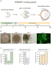
Figure 5. STEM-RET Scoring System of Retinal Differentiation from Stem Cells (SC)
A) Diagram of the three key stages of retinal development scored in the STEM-RET protocol (eye field specification, optic cup formation and retinal differentiation). The corresponding growth conditions (KSR: ;MM1: ;MM2: +RA: +taurine) and timing (days) are indicated below each stage for mouse iPSCs or ESCs. B) Representative micrographs of spheres from SC corresponding to days 1, 3 and 7 of STEM-RET using bright-field light microscopy. The outcroppings at day 7 (arrows) represent retinal tissue as indicated by the expression of GFP from the Rx-GFP transgenic EB5 mouse ESC line. The right panel for day 7 shows GFP expression alone. Scale bar, 200 mm. C) Representative examples of STEM-RET scoring of spheres of retinal differentiation for a single mouse SC line are shown. Eye field scoring: green; optic cup scoring: blue; and retinal differentiation: orange. Each bar represents the mean and standard deviation of integrate scores for each parameter on day 7 (eye field), day 10 (optic cup) and day 28 (retinal differentiation). Gray background bars: score for the positive control EB5 ESC line. All scores are normalized to normal mouse retina (score of 1.0). EFE, eye field efficiency; EFS, eye field specificity; EFP, eye field proliferation; OCE, optic cup efficiency; OCF, optic cup frequency; RD Q, retinal differentiation Q-PCR; RDEM, retinal differentiation electron microscopy; RDIF; retinal differentiation immunofluorescence.

Similar articles

Cited by

References

    1. Rodriguez-Galindo C, et al. Retinoblastoma. In: Orkin S, editor. Hematology and Oncology of Infancy and Childhood. Elsevier; 2015. pp. 1747–1778.
    1. Rodriguez-Galindo C, et al. Retinoblastoma: one world, one vision. Pediatrics. 2008;122:e763–770. - PMC - PubMed
    1. Zhang J, et al. A novel retinoblastoma therapy from genomic and epigenetic analyses. Nature. 2012;481:329–334. - PMC - PubMed
    1. Benavente CA, et al. Cross-species genomic and epigenomic landscape of retinoblastoma. Oncotarget. 2013;4:844–859. - PMC - PubMed
    1. McEvoy J, et al. RB1 gene inactivation by chromothripsis in human retinoblastoma. Oncotarget. 2014;5:438–450. - PMC - PubMed

LinkOut - more resources