. 2017 Feb 1;81(3):193-202.
doi: 10.1016/j.biopsych.2016.06.008. Epub 2016 Jun 16.

Sociability Deficits and Altered Amygdala Circuits in Mice Lacking Pcdh10, an Autism Associated Gene

Affiliations

Sociability Deficits and Altered Amygdala Circuits in Mice Lacking Pcdh10, an Autism Associated Gene

Hannah Schoch et al. Biol Psychiatry. .

Abstract

Background: Behavioral symptoms in individuals with autism spectrum disorder (ASD) have been attributed to abnormal neuronal connectivity, but the molecular bases of these behavioral and brain phenotypes are largely unknown. Human genetic studies have implicated PCDH10, a member of the δ2 subfamily of nonclustered protocadherin genes, in ASD. PCDH10 expression is enriched in the basolateral amygdala, a brain region implicated in the social deficits of ASD. Previous reports indicate that Pcdh10 plays a role in axon outgrowth and glutamatergic synapse elimination, but its roles in social behaviors and amygdala neuronal connectivity are unknown. We hypothesized that haploinsufficiency of Pcdh10 would reduce social approach behavior and alter the structure and function of amygdala circuits.

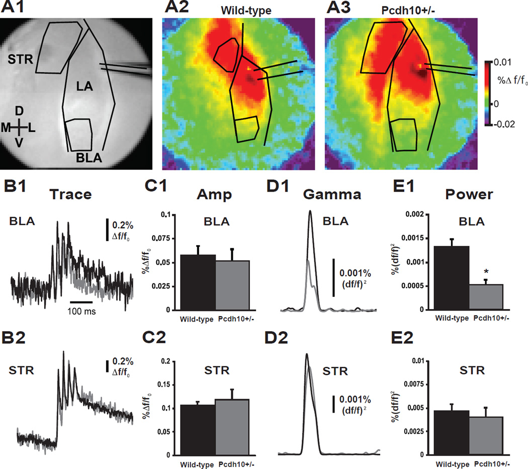
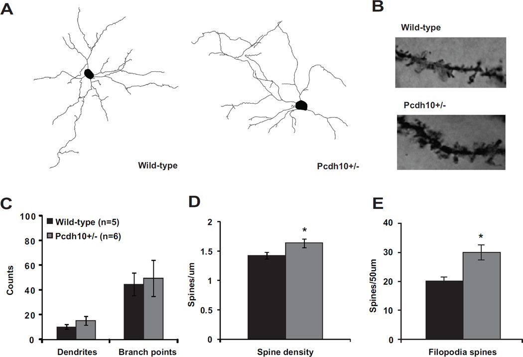
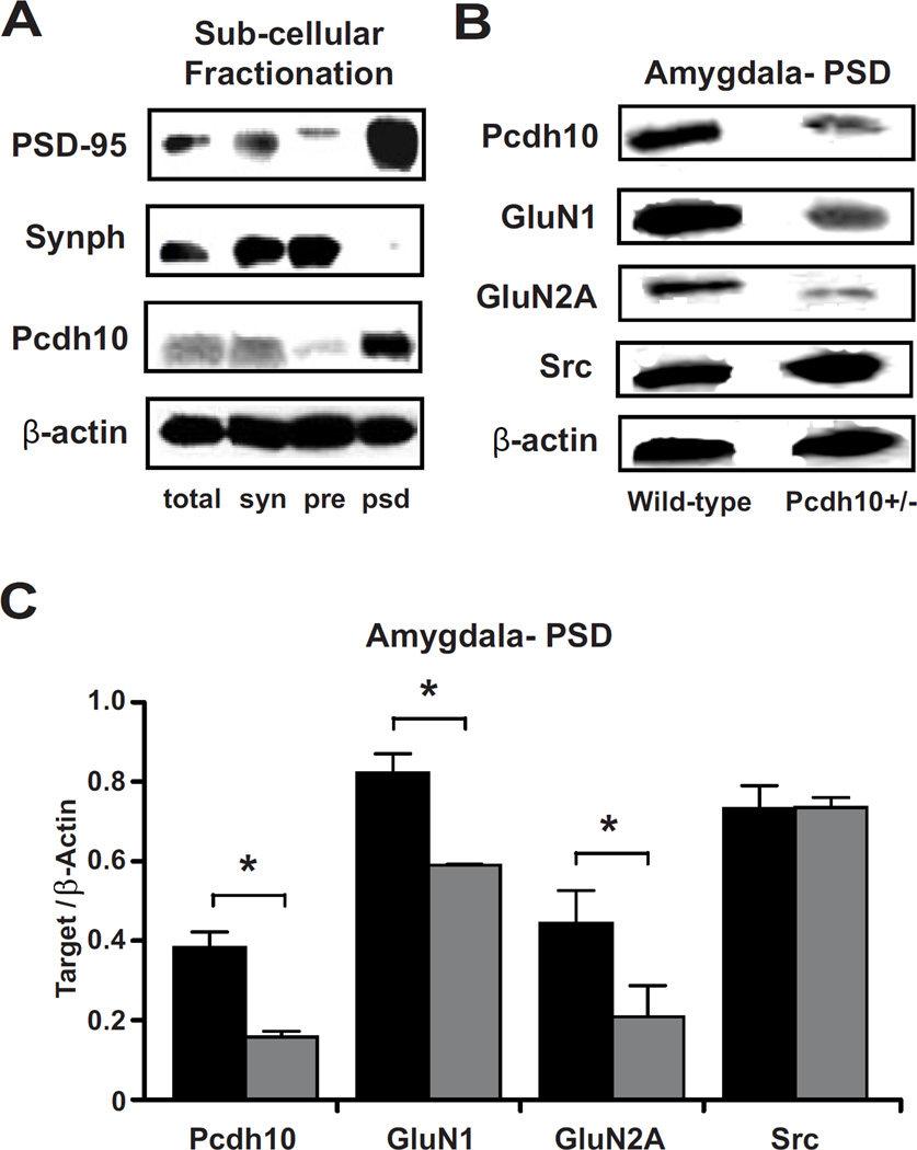
Methods: Mice lacking one copy of Pcdh10 (Pcdh10+/-) and wild-type littermates were assessed for social approach and other behaviors. The lateral/basolateral amygdala was assessed for dendritic spine number and morphology, and amygdala circuit function was studied using voltage-sensitive dye imaging. Expression of Pcdh10 and N-methyl-D-aspartate receptor (NMDAR) subunits was assessed in postsynaptic density fractions of the amygdala.

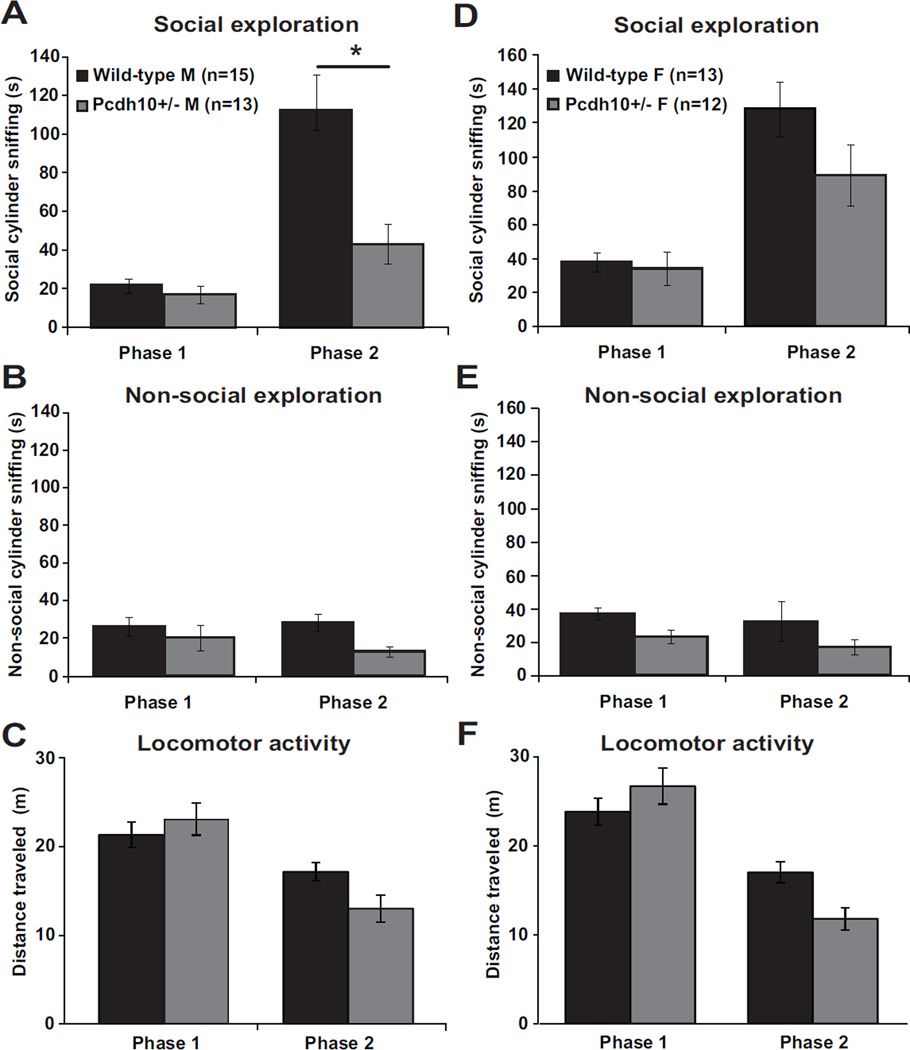
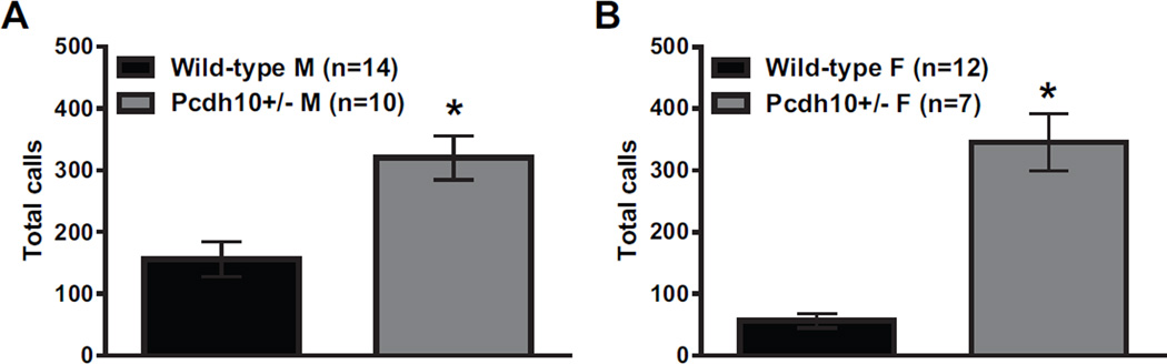
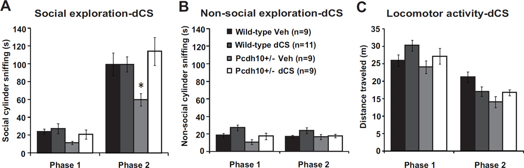
Results: Male Pcdh10+/- mice have reduced social approach behavior, as well as impaired gamma synchronization, abnormal spine morphology, and reduced levels of NMDAR subunits in the amygdala. Social approach deficits in Pcdh10+/- male mice were rescued with acute treatment with the NMDAR partial agonist d-cycloserine.

Conclusions: Our studies reveal that male Pcdh10+/- mice have synaptic and behavioral deficits, and establish Pcdh10+/- mice as a novel genetic model for investigating neural circuitry and behavioral changes relevant to ASD.

Keywords: Amygdala; Autism; Gene; NMDA; Protocadherin; Synapse.


Conflict of interest statement

Financial Disclosures Warren Bilker has consulted for Janssen Pharmaceuticals. The consulting is not related to the subject matter of the manuscript. Robert T. Schultz has received consulting fees from Akili Inc, Johnson & Johnson Consumer Inc, and Lumos Pharma Inc. The consulting is not related to the subject matter of the manuscript. All other authors report no biomedical financial interests or potential conflicts of interest.

Figures

Figure 1
Figure 1. Reduced social approach behavior in juvenile Pcdh10+/− mice
a) Time spent sniffing the social cylinder in male Pcdh10+/− and WT littermates in Phase 1 (stimulus mouse absent) and Phase 2 (stimulus mouse present). The raw data are shown in the graph, but a log transformation was required for normality in order to carry out data analysis. Male Pcdh10+/− mice showed lower sociability, i.e., significantly less increase in social cylinder sniffing duration from Phase 1 (absence of stimulus mouse) to Phase 2 (presence of stimulus mouse), than did male WT littermates (phase by genotype interaction, p=0.037, Figure 1a). There was a significant main effect of phase (p=0.001), but not a significant main effect of genotype across both phases (p=0.067). b) Time spent sniffing the non-social cylinder (which was empty in Phase 1 and had an object in Phase 2) in male mice. The raw data are shown in the graph, but a log transformation was required for normality in order to carry out data analysis. There was no significant phase by genotype interaction (p=0.612), main effect of phase (p=0.238), or main effect of genotype (p=0.180). c) Distance traveled by male mice. There was a significant phase by genotype interaction (p<0.001), with a significant main effect of phase (p<0.001) but no significant main effect of genotype (p=0.121). d) Time spent sniffing the social cylinder in female Pcdh10+/− and WT littermates in Phase 1 and Phase 2. The raw data are shown in the graph, but a log transformation was required for normality in order to carry out data analysis. There was no significant phase by genotype interaction (p=0.583), but there was a main effect of phase (p=0.001) and a significant main effect of genotype across both phases (p=0.039). e) Time spent sniffing the nonsocial cylinder in female mice. The raw data are shown in the graph, but a log transformation was required for normality in order to carry out data analysis. There was no significant phase by genotype interaction (p=0.888), but there were significant main effects of phase (p=0.006) and genotype (p=0.022). f) Distance traveled by female mice. There was a significant phase by genotype interaction (p<0.001), a significant main effect of phase (p<0.001), but no significant main effect of genotype (p=0.167).
Figure 2
Figure 2. Increased ultrasonic vocalizations (USVs) in Pcdh10+/− pups
Analysis was performed in Avisoft SASLab Pro (version 5.2.09; Avisoft Bioacoustics) with the following settings: 1024 FFT length, 100% frame, Hamming window, 75% time window overlap, 15 and 500 kHz frequency cutoffs, amplitude threshold of −60 dB, and a hold time of 30ms (78). a) Male Pcdh10+/− mice showed significantly increased numbers of USVs relative to male WT littermates (effect of genotype p<0.001) over the 5min recording period on postnatal day 6. b) Female Pcdh10+/− mice also showed significantly increased numbers of USVs relative to female WT littermates (effect of genotype p<0.001) (Figure 2) over 5min.
Figure 3
Figure 3. Pcdh10+/− mice exhibit BLA-specific impairment for the transmission of gamma-band power, but not amplitude of EPSP
a1) Grey scale image of amygdala coronal slice showing the lateral amygdala (LA), basolateral amygdala (BLA) and striatum (STR), electrode placement and regions of interest (ROI). Color images show peak VSDi responses following direct stimulation of the LA in slices from wild-type (a2) and Pcdh10+/− (a3) males. Data were displayed as the change in fluorescence divided by the resting fluorescence (Δf/f0). Depolarizing Δf/f0 signals were displayed as warmer colors and hyperpolarizations were represented as colder colors. Evoked average VSDi signal responses in the BLA (b1) and STR (b2) to 4 stimuli at 40Hz over time are shown. Traces from wild-type mice are shown in black and Pcdh10+/− are shown in gray. Statistical comparisons were made using a Mann-Whitney test. No differences were seen in the peak amplitude response in BLA (c1) or STR (c2) between wild-type and Pcdh10+/− (n=5; BLA, p=0.754; STR, p=0.754). In contrast, taking the integral of the gamma-band response (30-50Hz) over time in both BLA (d1) and STR (d2) ROIs, revealed specific reduction in the gamma band coherence of the BLA in response to high frequency LA stimulation (BLA, p=0.016; STR p=0.917). This was specific to the LA - BLA circuit (e1), as functional connectivity to the striatum (e2) remained unaffected.
Figure 4
Figure 4. Increased filopodia-type spines on lateral/basolateral amygdala neurons of Pcdh10+/− males
a) Representative dendritic reconstructions from lateral/basolateral (LA/BLA) amygdala neurons from wild-type and Pcdh10+/− males. b) Representative dendritic lengths from LA/BLA neurons from wild-type and Pcdh10+/− males. c) Counts of dendrites and branch points in LA/BLA of wild-type and Pcdh10+/− males. A mixed model was used to compare the mean dendrites and branch points between the two genotypes, while considering the non-independence of multiple measurements within some mice. There was no difference in dendrites (p=0.190) or branch points (p=0.771) between the genotypes. d) A mixed model was used to compare the mean spine density in the LA/BLA between the two genotypes, while considering the non-independence of multiple measurements within some mice. Based on the available literature on the role of Pcdh10 in spine elimination (17), a one-tailed test was applied to test our a priori directional hypothesis. Indeed, Pcdh10+/− mice had significantly higher spine density that WT littermates (p=0.048). e) A mixed model was fit separately for each spine type with a one-sided test, based on the a priori hypothesis of higher spine counts in Pcdh10+/− mice (17). Relative to WT, Pcdh10+/− amygdala neurons showed significantly higher levels of filopodia spines (p=0.030)
Figure 5
Figure 5. Decreased Pcdh10 in the PSD of Pcdh10+/− males was associated with decreased GLUN1 in the PSD
a) Pcdh10 was found to be concentrated in the post-synaptic density fraction (psd) where PSD-95, the marker for PSD is most concentrated; whereas it was found to be poorly represented in presynaptic fraction (pre) where Synaptophysin (Synph), the presynaptic marker, is heavily concentrated. b) Representative blots of Pcdh10, GluN1, GluN2A and β-actin in the PSD fractions of wild-type and Pcdh10+/− mice showing decreased Pcdh10, GluN1, and GluN2A in the amygdala of Pcdh10+/− mice. c) Pcdh10+/− mice showed significantly reduced levels of Pcdh10 (p<0.001), GluN1 (p<0.001), and GluN2A (p=0.011), but not SRC (p=0.939).
Figure 6
Figure 6. NMDAR agonist d-cycloserine rescued sociability impairment in Pcdh10+/− males
a) Treatment with acute d-cycloserine (dCS 32mg/kg) increased social exploration in juvenile Pcdh10+/− males. The raw data are shown in the graph, but a log transformation was required for normality in order to carry out data analysis. A mixed model was fit including the effects of phase (1,2), genotype (Pcdh10+/−, WT), and drug (dCS, vehicle) on social cylinder sniffing. There was a significant phase by genotype interaction (p=0.020), but no other significant 2-way interactions. There were significant main effects of phase (p<0.001), genotype (p<0.001), and drug (p=0.019). To help interpret the genotype and drug effects, a separate model was fit for phase 2 only. In this model, there was a significant genotype by drug interaction (p=0.020) with significant main effects of genotype (p=0.006) and drug (p=0.001). b) Treatment with dCS and non-social cylinder sniffing. The raw data are shown in the graph, but a log transformation was required for normality in order to carry out data analysis. In a mixed model including phase, genotype, and drug, there was a significant phase by genotype interaction (p=0.020) but no other significant interactions. There were significant main effects of phase (p=0.009), genotype (p<0.001), and drug (p=0.026). To help interpret the genotype and drug effects, a separate model was fit for phase 2 only. In this model, there was no significant genotype by drug interaction (p=0.730) and no significant main effects of genotype (p=0.251) or drug (p=0.217). c) Treatment with dCS and distance traveled. In a mixed model including phase, genotype, and drug, there was a significant three-way interaction of phase by genotype by drug (p=0.046), but no significant two-way interactions. There was also a significant main effect of phase (p<0.001), but no main effects of genotype (p=0.386) or drug (0.158) on distance traveled. To help interpret the of genotype and drug effects, a separate model was fit for phase 2 only, which found a significant genotype by drug interaction (p=0.009) with a main effect of genotype (<0.001) but not drug (p=0.156).

Comment in

  • Protocadherins and the Social Brain.
    Tsai NP, Huber KM. Tsai NP, et al. Biol Psychiatry. 2017 Feb 1;81(3):173-174. doi: 10.1016/j.biopsych.2016.10.025. Biol Psychiatry. 2017. PMID: 28024704 Free PMC article. No abstract available.

References

    1. Chevallier C, Kohls G, Troiani V, Brodkin ES, Schultz RT. The social motivation theory of autism. Trends in Cognitive Sciences. 2012:231–238. - PMC - PubMed
    1. Werling DM, Geschwind DH. Sex differences in autism spectrum disorders. Curr Opin Neurol. 2013 Apr;26(2):146–153. - PMC - PubMed
    1. Hartley SL, Sikora DM. Sex differences in Autism spectrum disorder: An examination of developmental functioning, Autistic symptoms, and coexisting behavior problems in toddlers. J Autism Dev Disord. 2009 Dec;39(12):1715–1722. - PMC - PubMed
    1. Gilman SR, Iossifov I, Levy D, Ronemus M, Wigler M, Vitkup D. Rare De Novo Variants Associated with Autism Implicate a Large Functional Network of Genes Involved in Formation and Function of Synapses. Neuron. 2011;70(5):898–907. - PMC - PubMed
    1. Irwin SA, Patel B, Idupulapati M, Harris JB, Crisostomo RA, Larsen BP, et al. Abnormal dendritic spine characteristics in the temporal and visual cortices of patients with fragile-X syndrome: A quantitative examination. Am J Med Genet. 2001;98(2):161–167. - PubMed

MeSH terms