Review
. 2017;57(4):975-999.
doi: 10.3233/JAD-160612.

Amyloid-Beta and Phosphorylated Tau Accumulations Cause Abnormalities at Synapses of Alzheimer's disease Neurons

Affiliations
Review

Amyloid-Beta and Phosphorylated Tau Accumulations Cause Abnormalities at Synapses of Alzheimer's disease Neurons

Ravi Rajmohan et al. J Alzheimers Dis. 2017.

Abstract

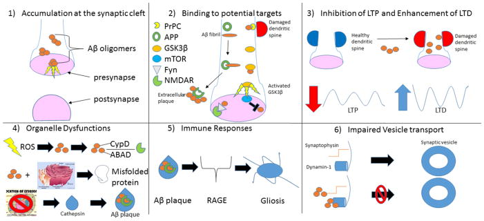
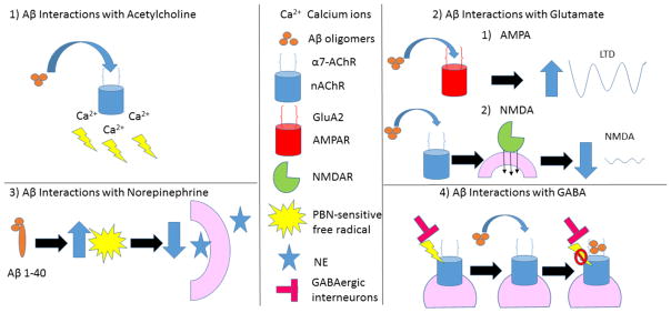
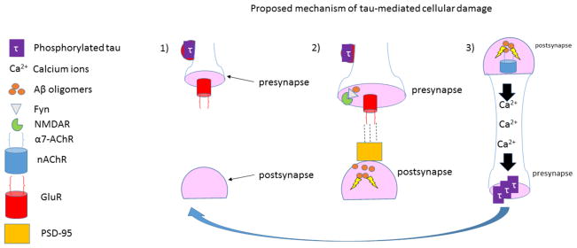
Amyloid-beta (Aβ) and hyperphosphorylated tau are hallmark lesions of Alzheimer's disease (AD). However, the loss of synapses and dysfunctions of neurotransmission are more directly tied to disease severity. The role of these lesions in the pathoetiological progression of the disease remains contested. Biochemical, cellular, molecular, and pathological studies provided several lines of evidence and improved our understanding of how Aβ and hyperphosphorylated tau accumulation may directly harm synapses and alter neurotransmission. In vitro evidence suggests that Aβ and hyperphosphorylated tau have both direct and indirect cytotoxic effects that affect neurotransmission, axonal transport, signaling cascades, organelle function, and immune response in ways that lead to synaptic loss and dysfunctions in neurotransmitter release. Observations in preclinical models and autopsy studies support these findings, suggesting that while the pathoetiology of positive lesions remains elusive, their removal may reduce disease severity and progression. The purpose of this article is to highlight the need for further investigation of the role of tau in disease progression and its interactions with Aβ and neurotransmitters alike.

Keywords: Amyloid; neurotransmitters; synapse; tau.


Figures

Figure 1
Figure 1
Amyloid beta’s many sites of neuronal damage. 1) Soluble intracellular oligomers of Aβ are cytotoxic [122,128]. 2) Interaction with PrPC leads to dendritic spine damage [139] (top right). Aβ fibril binds to APP during formation of extracellular plaques [144] (middle left). Aβ inappropriately activates GSK3β [105] (middle right). Aβ synergizes with Fyn to disrupt synaptic receptors [104] (bottom left). mTOR may inhibit Aβ [105] (bottom right). 3) Mounting evidence suggests Aβ-mediated destruction of spines and synapses leads to LTP impairment and LTD enhancement [105]. 4) Mitochondrial distress may lead to production of ROS that may accelerate Aβ formation. Aβ may then affect NMDAR activity by binding to intracellular mitochondrial CypD or mitochondrial ABAD [130] (Top). Aβ-mediated cytotoxicity may be particularly damaging to the endoplasmic reticulum, affecting its production and modification of crucial proteins [234] (middle). Identification of cathepsins in Aβ plaques suggest protease dysfunctions from lysosomes may play a critical role in advancement of pathology [235] (bottom). 5) RAGE may play a prominent role in the increased microglial activation and proinflammatory markers commonly associated with senile plaques [160]. 6) Aβ interacts with synaptophysin in presynaptic terminals of the hippocampus [256] to impair neurotransmission (top). Aβ also interferes with dynamin-1 allowing synaptic vesicles to reenter the synaptic pool [257,258] (bottom).
Figure 2
Figure 2
Summary of amyloid beta’s interaction with neurotransmitters at neuronal synapses. 1) Aβ may lead to an overactivation of nAChRs and excessive Ca2+ influx that impairs the signaling cascades of memory formation [16, 25]. 2) Aβ oligomers are believed to interact with the GluA2 subunit of AMPAR [274] leading to increased LTD (top). Aβ promotes endocytosis of NMDARs in cortical neurons and produces a rapid and persistent depression of NMDA-evoked currents via activation of nAChRs [138, 280, 281] (bottom). 3) Aβ1–40 allows a PBN-sensitive free radical to deleteriously effect evoked NE release [285]. Aβ inhibits nicotinic currents from GABAergic interneurons by directly blocking the postsynaptic a7-nAChR channels [263].
Figure 3
Figure 3
Tau at the synapse. 1) Phosphorylated tau accumulates in dendritic spines where it may affect the synaptic trafficking and anchoring of GluR [296]. 2) Dendritic tau-mediated Fyn recruitment and GluR2/PSD-95 interaction confer Aβ toxicity at the postsynapse. 3) Upon activation by Aβ, a7 AChR mediates a rise in intracellular Ca2+, which may activate kinases that phosphorylate tau [328, 329]. Thus, Aβ and hyperphosphorylated tau may promote each other’s role in the pathogenesis of Alzheimer’s disease.

References

    1. DeKosky ST, Scheff SW. Synapse loss in frontal cortex biopsies in Alzheimer disease: correlation with cognitive severity. Annals of neurology. 1990;27(5):457–464. - PubMed
    1. Scheff SW, DeKosky ST, Price DA. Quantitative assessment of cortical synaptic density in Alzheimer disease. Neurobiology of aging. 1990;11(1):29–37. - PubMed
    1. Scheff SW, Price DA. Synapse loss in the temporal lobe in Alzheimer disease. Annals of neurology. 1993;33(2):190–199. - PubMed
    1. Scheff SW, Price DA, Schmitt FA, DeKosky ST, Mufson EJ. Synaptic alterations in CA1 in mild Alzheimer disease and mild cognitive impairment. Neurology. 2007;68(18):1501–1508. - PubMed
    1. Terry RD, Masliah E, Salmon DP, Butters N, DeTeresa R, Hill R, Hansen LA, Katzman R. Physical basis of cognitive alterations in Alzheimer disease: synapse loss is the major correlate of cognitive impairment. Ann Neurol. 1991;30(4):572–580. doi: 10.1002/ana.410300410. - DOI - PubMed