. 2016 Oct;468(10):1685-95.
doi: 10.1007/s00424-016-1873-y. Epub 2016 Aug 27.

In vivo definition of cardiac myosin-binding protein C's critical interactions with myosin

Affiliations

In vivo definition of cardiac myosin-binding protein C's critical interactions with myosin

Md Shenuarin Bhuiyan et al. Pflugers Arch. 2016 Oct.

Abstract

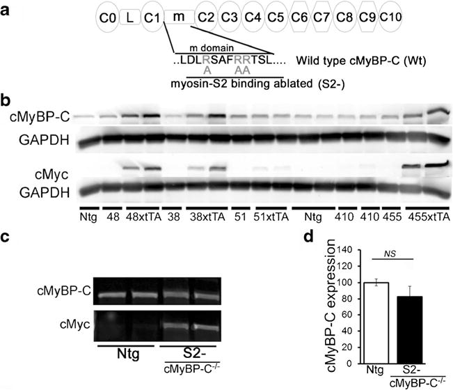
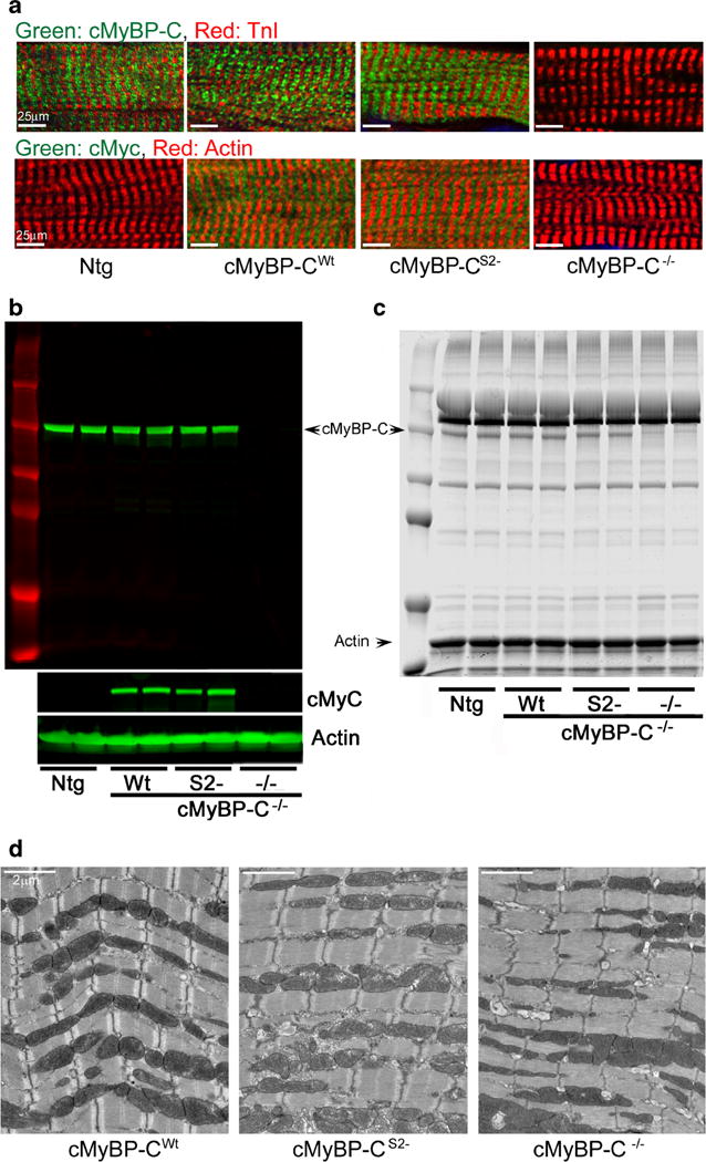
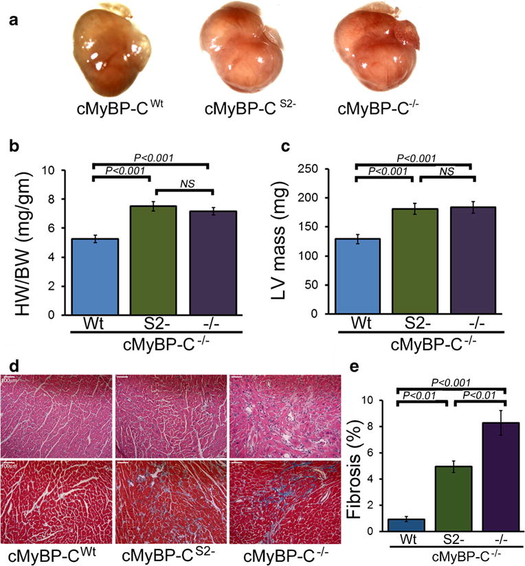
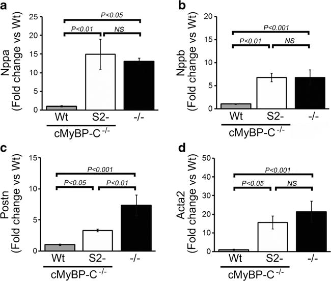
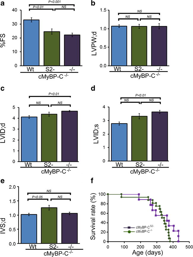
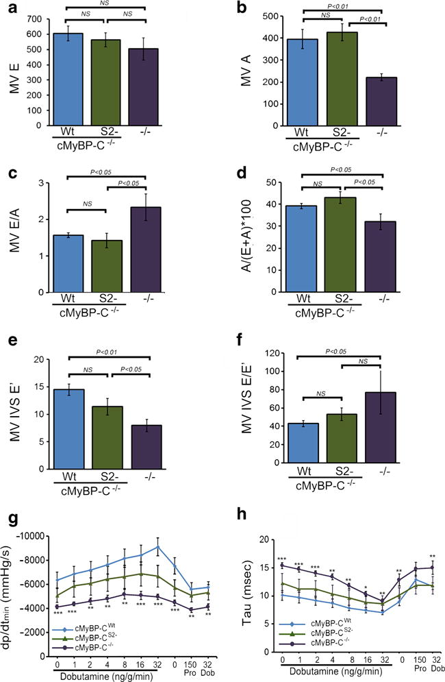
Cardiac myosin-binding protein C (cMyBP-C) is an integral part of the sarcomeric machinery in cardiac muscle that enables normal function. cMyBP-C regulates normal cardiac contraction by functioning as a brake through interactions with the sarcomere's thick, thin, and titin filaments. cMyBP-C's precise effects as it binds to the different filament systems remain obscure, particularly as it impacts on the myosin heavy chain's head domain, contained within the subfragment 2 (S2) region. This portion of the myosin heavy chain also contains the ATPase activity critical for myosin's function. Mutations in myosin's head, as well as in cMyBP-C, are a frequent cause of familial hypertrophic cardiomyopathy (FHC). We generated transgenic lines in which endogenous cMyBP-C was replaced by protein lacking the residues necessary for binding to S2 (cMyBP-C(S2-)). We found, surprisingly, that cMyBP-C lacking the S2 binding site is incorporated normally into the sarcomere, although systolic function is compromised. We show for the first time the acute and chronic in vivo consequences of ablating a filament-specific interaction of cMyBP-C. This work probes the functional consequences, in the whole animal, of modifying a critical structure-function relationship, the protein's ability to bind to a region of the critical enzyme responsible for muscle contraction, the subfragment 2 domain of the myosin heavy chain. We show that the binding is not critical for the protein's correct insertion into the sarcomere's architecture, but is essential for long-term, normal function in the physiological context of the heart.

Keywords: Cardiac; Heart; Myofilament; Myosin; Myosin-binding protein C; Sarcomere.


Figures

Fig. 1
Fig. 1
Expression of inducible, mutated cMyBP-C protein. a Schematic diagram showing domain organization and sequences of wild-type cMyBP-C (Wt) and cMyBP-CS2− (m domain; R266, R270, R271 mutated to alanine). b Expression of different lines of the S2- transgene in the S2-xtTA mouse hearts. Representative Western blots using anti-cMyBP-C and anti-c-myc antibodies. α-Sarcomeric actin was used as a loading control. c Western blot analyses of cMyBP-C levels in the heart. To obtain complete replacement of endogenous cMyBP-C with the Tg mutant proteins, the individual cMyBP-CS2−-xtTA double Tg lines were bred into the cMyBP-C−/− background. Transgenic expression of the cMyBP-CS2− protein approximated normal levels of the protein in the Ntg hearts. d Quantitation of transgene expression via Western blotting confirmed equivalent expression of cMyBP-CS2− protein relative to normal cMyBP-C protein in the Ntg hearts
Fig. 2
Fig. 2
Expression and sarcomere incorporation of cMyBP-CS2−. The S2 − and Wt constructs were each bred into the cMyBP-C−/− backgrounds. a Expression and sarcomere incorporation of the c-myc-tagged, Tg cMyBP-C in the cMyBP-CWt, cMyBP-CS2−, and cMyBP-C−/− mice defined by immunofluorescent staining with anti-cMyBP-C (green) and cardiac TnI antibody (red). All Tg samples were derived from 12-week-old hearts. b Representative Western blots derived from myofilament protein preparations. α-Sarcomeric actin was used as a loading control. c SDS-PAGE analyses of myofibrillar proteins from Ntg, cMyBP-CWt, cMyBP-CS2−, and cMyBP-C−/− hearts. d Transmission electron micrographs of the sarcomeres
Fig. 3
Fig. 3
Characterization of the cMyBP-CS2− hearts at 3 months. The S2− and Wt constructs were each bred into the cMyBP-C−/− backgrounds. a Hearts from cMyBP-CWt, cMyBP-CS2−, and cMyBP-C−/− mice. b Heart weight/body weight ratios. c Changes in LV mass index determined by echocardiography (n ≥ 6 mice per group). d H&E (top) and Masson’s trichrome (bottom) staining of cardiac sections. e Blue-stained areas were color-thresholded using NIH ImageJ software and quantitated as described in “Methods.” All analyses were carried out with 3-month-old mice. Scale bars 100 μm
Fig. 4
Fig. 4
Expression of markers denoting cardiac stress. a, b Nppa and Nppb transcript levels. Values are expressed as fold change versus cMyBP-CWt control (n = 6 per group). c, d mRNA expression of Postn and Acta2. Values are expressed as fold change versus cMyBP-CWt (n = 6 per group). All samples were derived from 3-month-old hearts (n = 4). P values were determined by Tukey’s post hoc test
Fig. 5
Fig. 5
M-mode echocardiography indices of LV end-diastolic diameter and function. a Percent fractional shortening (%FS). b LV diastolic posterior wall thickness. c LV diastolic volume (LV vol;d). d LV systolic volume (LV vol;s). e Diastolic interventricular septum thickness (IVS;d). (n ≧ 10 mice per group, mean ± S.E.M). P value versus cMyBP-CWt mice by Tukey’s post hoc test. All measurements were carried out with 3-month-old mice. f Kaplan-Meier analysis of survival probabilities for the null (n = 16) versus the cMyBP-CS2− (n = 9) animals. There were no statistically significant differences between the two curves
Fig. 6
Fig. 6
Diastolic function. a, b Doppler echocardiographic measurements of mitral inflow velocity during early diastolic filling (E wave) and mitral inflow velocity during atrial contraction (A wave). c Absolute values for early-to-late diastolic flow velocity ratio (E/A), used as an index of LV diastolic filling. d Contribution (in percent) of atrial contraction to diastolic filling (n ≥ 6 mice per group, values are means ± SEM). e Tissue Doppler measurements of early diastolic mitral annular velocities (E′) at the septal (IVS) wall. f Relationship between early diastolic inflow velocity and early diastolic tissue velocity (E/E′) at the septal (IVS) wall (n ≥ 6 mice per group, values are means ± SEM). g, h Maximum rate of LVrelaxation (dP/dtmin and relaxation time constant (tau) in 4.5-month-old cMyBP-CWt, cMyBP-CS2−, and cMyBP-C−/− mice during closed-chest cardiac catheterization, measured at baseline and with increasing adrenergic stimulation via dobutamine infusion. Propranolol was then delivered to produce complete β-adrenergic blockade (n ≥ 6 mice per group, values are means ± SEM). *P < 0.05 and ***P < 0.001 versus cMyBP-CWt animals

References

    1. Bezold KL, Shaffer JF, Khosa JK, Hoye ER, Harris SP. A gain-of-function mutation in the M-domain of cardiac myosin-binding protein-C increases binding to actin. J Biol Chem. 2013;288:21496–21505. doi: 10.1074/jbc.M113.474346. - DOI - PMC - PubMed
    1. Bhuiyan MS, Gulick J, Osinska H, Gupta M, Robbins J. Determination of the critical residues responsible for cardiac myosin binding protein C’s interactions. J Mol Cell Cardiol. 2012;53:838–847. doi: 10.1016/j.yjmcc.2012.08.028. - DOI - PMC - PubMed
    1. Bhuiyan MS, Pattison JS, Osinska H, James J, Gulick J, McLendon PM, Hill JA, Sadoshima J, Robbins J. Enhanced autophagy ameliorates cardiac proteinopathy. J Clin Invest. 2013;123:5284–5297. doi: 10.1172/JCI70877. - DOI - PMC - PubMed
    1. Carrier L, Bonne G, Bahrend E, Yu B, Richard P, Niel F, Hainque B, Cruaud C, Gary F, Labeit S, Bouhour JB, Dubourg O, Desnos M, Hagege AA, Trent RJ, Komajda M, Fiszman M, Schwartz K. Organization and sequence of human cardiac myosin binding protein C gene (MYBPC3) and identification of mutations predicted to produce truncated proteins in familial hypertrophic cardiomyopathy. Circ Res. 1997;80:427–434. - PubMed
    1. Carrier L, Knoll R, Vignier N, Keller DI, Bausero P, Prudhon B, Isnard R, Ambroisine ML, Fiszman M, Ross J, Jr, Schwartz K, Chien KR. Asymmetric septal hypertrophy in heterozygous cMyBP-C null mice. Cardiovasc Res. 2004;63:293–304. doi: 10.1016/j.cardiores.2004.04.009. - DOI - PubMed

Publication types

LinkOut - more resources