. 2017 Jan;137(1):85-94.
doi: 10.1016/j.jid.2016.07.039. Epub 2016 Aug 26.

Spread of Psoriasiform Inflammation to Remote Tissues Is Restricted by the Atypical Chemokine Receptor ACKR2

Affiliations

Spread of Psoriasiform Inflammation to Remote Tissues Is Restricted by the Atypical Chemokine Receptor ACKR2

Kave Shams et al. J Invest Dermatol. 2017 Jan.

Abstract

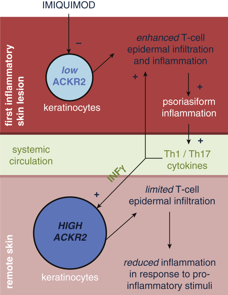
Elucidating the poorly defined mechanisms by which inflammatory lesions are spatially restricted in vivo is of critical importance in understanding skin disease. Chemokines are the principal regulators of leukocyte migration and are essential in the initiation and maintenance of inflammation. The membrane-bound psoriasis-associated atypical chemokine receptor 2 (ACKR2) binds, internalizes and degrades most proinflammatory CC-chemokines. Here we investigate the role of ACKR2 in limiting the spread of cutaneous psoriasiform inflammation to sites that are remote from the primary lesion. Circulating factors capable of regulating ACKR2 function at remote sites were identified and examined using a combination of clinical samples, relevant primary human cell cultures, in vitro migration assays, and the imiquimod-induced model of psoriasiform skin inflammation. Localized inflammation and IFN-γ together up-regulate ACKR2 in remote tissues, protecting them from the spread of inflammation. ACKR2 controls inflammatory T-cell chemotaxis and positioning within the skin, preventing an epidermal influx that is associated with lesion development. Our results have important implications for our understanding of how spatial restriction is imposed on the spread of inflammatory lesions and highlight systemic ACKR2 induction as a therapeutic strategy in the treatment and prevention of psoriasis and potentially a broad range of other immune-mediated diseases.


Figures

Figure 1
Figure 1
ACKR2-deficient mice display an exaggerated psoriasiform phenotype in response to IMQ treatment. (a) WT mice treated for 3 days (culled day 4) with (top) vehicle control and (middle) IMQ; (bottom) IMQ-treated ACKR2-deficient mice. Scale bars = 1 cm. (b) Hematoxylin and eosin-stained WT and ACKR2-deficient (KO) skin after 3 days of IMQ treatment and quantification of epidermal thickness after 3 or 6 days of daily IMQ. Scale bars = 100 μm. Statistics: one-way analysis of variance. (c) Ki67-stained WT and ACKR2-deficient (KO) skin after 3 days of IMQ and quantification of thickness of Ki67 staining in the epidermis. (d) (top) PASI skin inflammation and (bottom) weight change (baseline = 100%) throughout treatments. Statistics: two-way analysis of variance, Tukey’s multiple comparisons test. (e) Correlation between mean epidermal thickness and PASI score (left). Correlation between mean thickness of Ki67 positive epidermis and PASI score (right). P < 0.05, ∗∗P < 0.01. ACKR2, atypical chemokine receptor 2; IMQ, imiquimod; KO, knockout; NS, not significant; PASI, Psoriasis Area Severity Index; WT, wild type.
Figure 2
Figure 2
ACKR2 regulates T-cell migration and positioning in the skin. (a) CD3+ staining after 3 days of treatment with IMQ. Arrows: CD3+ T cells. Red line: dermal-epidermal junction. (b) Distance of CD3+ cells from basement membrane after 3 days of treatment with IMQ. Two-way analysis of variance, Sidak’s multiple comparisons test. (c) Epidermal CD3+ as percentage total CD3+ cells (one-way analysis of variance, Tukey’s multiple comparisons test). (d) T-cell migration toward CCL5 (top of plots) across low or high ACKR2-expressing keratinocytes. Plots on left: cumulative migration direction. Plots on right: individual tracks (black = toward and red = away from CCL5). Statistics: Rayleigh test. (e) ACKR2 expression in heart and liver from vehicle- (black bars) or IMQ- (grey bars) treated mice. (f) ACKR2 expression in draining lymph nodes from treated or untreated skin. Mice were treated 3 days with vehicle (black bars) or IMQ (grey bars). (g) ACKR2 at site of vehicle/IMQ treatment and distal dorsal skin. ∗∗P < 0.01, ∗∗∗P < 0.005, ∗∗∗∗P < 0.0001. ACKR2, atypical chemokine receptor 2; IMQ, imiquimod; KO, knockout; NS, not significant; WT, wild type.
Figure 3
Figure 3
Cytokines from activated T cells induce ACKR2 expression in human keratinocytes. ACKR2 expression in response to cytokines (100ng/ml) in (a) human keratinocytes, (b) skin equivalents treated with activated CD4+ T cells and (c) skin equivalents after 2–4 days with/without treatment with all-trans retinoic acid or cyclosporine A. ACKR2 expression in (d) human keratinocytes treated with T-cell conditioned medium alone and (e) human keratinocytes treated with anti–IFN-γ antibodies. Two-way analysis of variance, Tukey’s MCT. (f) Mouse IFN-γ expression 24 hours after treatment. Virus-infected samples act as positive control. One-way analysis of variance, Tukey’s multiple comparisons test. (g) ACKR2 expression in mouse dorsal skin after IFN-γ treatment (20,000U/day for 4 days, n = 6). Student t test. (h) WT mice were treated for 3 days and photographed on day 4. Scale bars = 1 cm. (i) PASI score. Arrows: first day of IFN-γ/phosphate buffered saline injections and IMQ/vehicle. (j) Correlation of cutaneous ACKR2 expression and PASI score. P < 0.05, ∗∗P < 0.01, ∗∗∗P < 0.005, ∗∗∗∗P < 0.0001. ACKR2, atypical chemokine receptor 2; CsA, cyclosporine A; d, days; IMQ, imiquimod; M, million; NS, not significant; PASI, Psoriasis Area Severity Index; RA, retinoic acid; TBP, TATA-binding protein; Th, t helper.
Figure 4
Figure 4
Localized IMQ combined with systemic IFN-γ ameliorates psoriasis-like pathology at both primary and distal sites in an ACKR2-dependent manner. (a) ACKR2 expression (day 5) in treated and distal/remote untreated skin of IMQ-treated mice (days 1–4) in presence/absence of systemic IFN-γ (20,000 U/day). Statistics: Student t test. (b) Experimental design. PASI score at (c) initial lesions and (d) lesion induced by IMQ at second site. (e) As in c and d with ear as second site. Ear thickness was measured in mice treated on the flank with IMQ and systemic IFN-γ (20,000 U/day) or PBS. (f) ACKR2 expression in second IMQ-induced lesions. First IMQ lesion was induced with concurrent PBS (grey bar) or IFN-γ (black bar). Statistics: Student t test. (g) ACKR2 expression in distal epidermis and dermis. Epidermal/dermal separation confirmed by involucrin quantitative PCR (grey bars). Statistics: Student t test. PASI score of IMQ-treated ACKR2-deficient mice pretreated with systemic PBS or IFN-γ (20,000 U/day) at (h) initial lesion and at (i) secondary lesion. Statistics: two-way analysis of variance. P < 0.05, ∗∗P < 0.01, ∗∗∗P < 0.005, ∗∗∗∗P < 0.0001. ACKR2, atypical chemokine receptor 2; IMQ, imiquimod; NS, not significant; PASI, Psoriasis Area Severity Index; PBS, phosphate buffered saline.
Figure 5
Figure 5
Histological and immunochemical analysis of primary and secondary IMQ-treated skin sites. (a, b) Representative images of hematoxylin and eosin, Ki67, and CD3+ staining and associated quantification of (a) initial IMQ-induced lesion with concurrent PBS or systemic IFN-γ (day 5) and (b) second IMQ-induced lesion initiated after cessation of initial IMQ/IFN-γ or PBS treatment (day 8) (as in Figure 4b). In CD3+ images, dark red arrows = epidermal CD3+ T cells and dark red line = dermal-epidermal junction. All scale bars = 100μm. (c–f) Total CD3+ T-cell numbers in IMQ-induced skin lesion with concurrent PBS or systemic IFN-γ in (c, d) first lesion or (e, f) second lesion after cessation of systemic PBS or IFN-γ treatment. Numbers shown are for (c, e) whole skin and (d, f) epidermis. Mean of three separate fields of view/mouse. P < 0.05, ∗∗P < 0.01, ∗∗∗P < 0.005, ∗∗∗∗P < 0.0001. ACKR2, atypical chemokine receptor 2; FOV, field of view; H&E, hematoxylin and eosin; IMQ, imiquimod; NS, not significant; PASI, Psoriasis Area Severity Index; PBS, phosphate buffered saline.
Figure 6
Figure 6
Schematic model of the main findings of the study. ACKR2, atypical chemokine receptor 2; Th, T helper.

Comment in

Similar articles

Cited by

References

    1. Bachelerie F., Ben-Baruch A., Burkhardt A.M., Combadiere C., Farber J.M., Graham G.J. International Union of Pharmacology. LXXXIX. Update on the extended family of chemokine receptors and introducing a new nomenclature for atypical chemokine receptors. Pharmacol Rev. 2014;66:1–9. - PMC - PubMed
    1. Bachelerie F., Graham G.J., Locati M., Mantovani A., Murphy P.M., Nibbs R. New nomenclature for atypical chemokine receptors. Nat Immunol. 2014;15:207–208. - PubMed
    1. Baldwin H.M., Pallas K., King V., Jamieson T., McKimmie C.S., Nibbs R.J.B. Microarray analyses demonstrate the involvement of Type I Interferons in psoriasiform pathology development in D6 deficient mice. J Biol Chem. 2013;288:36473–36483. - PMC - PubMed
    1. Bazzan E., Saetta M., Turato G., Borroni E.M., Cancellieri C., Baraldo S. Expression of the atypical chemokine receptor D6 in human alveolar macrophages in chronic obstructive pulmonary disease. Chest. 2012;143:98–106. - PubMed
    1. Borroni E.M., Cancellieri C., Vacchini A., Benureau Y., Lagane B., Bachelerie F. Beta-arrestin-dependent activation of the cofilin pathway is required for the scavenging activity of the atypical chemokine receptor D6. Sci Signal. 2013 6 ra30.1–11, S1–3. - PubMed

MeSH terms