. 2016 Sep 13;7(3):557-570.
doi: 10.1016/j.stemcr.2016.07.017. Epub 2016 Aug 25.

Expandable and Rapidly Differentiating Human Induced Neural Stem Cell Lines for Multiple Tissue Engineering Applications

Affiliations

Expandable and Rapidly Differentiating Human Induced Neural Stem Cell Lines for Multiple Tissue Engineering Applications

Dana M Cairns et al. Stem Cell Reports. .

Abstract

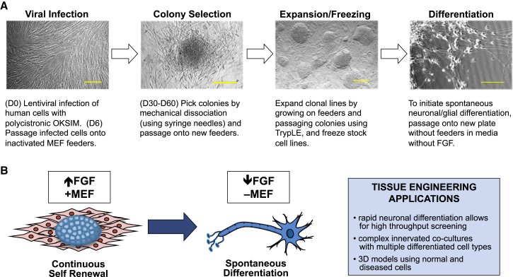
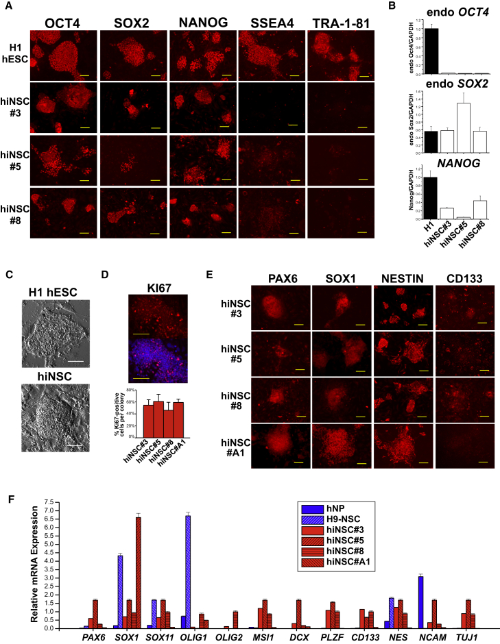
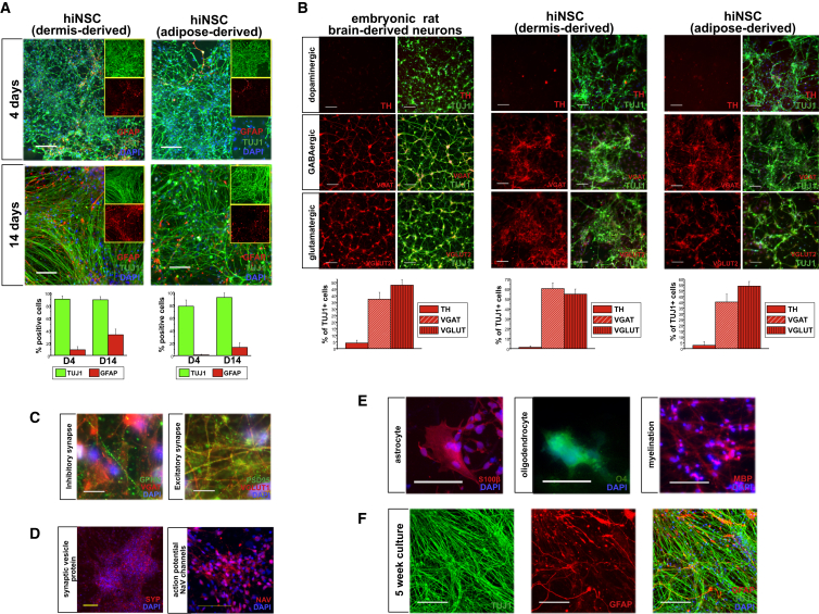
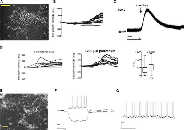
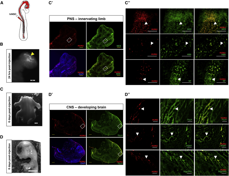
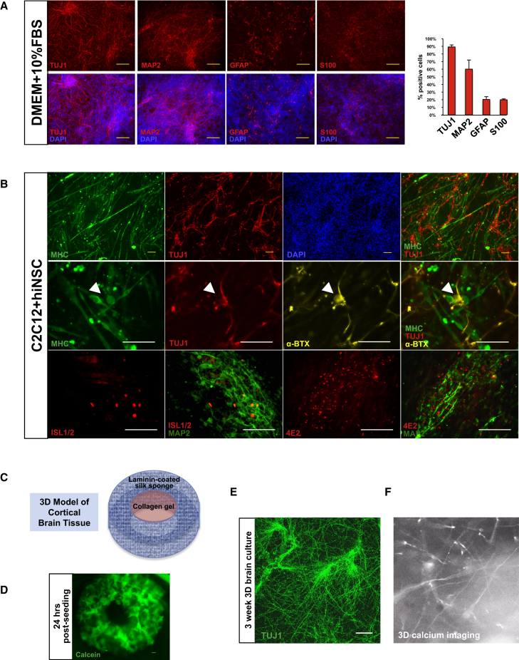
Limited availability of human neurons poses a significant barrier to progress in biological and preclinical studies of the human nervous system. Current stem cell-based approaches of neuron generation are still hindered by prolonged culture requirements, protocol complexity, and variability in neuronal differentiation. Here we establish stable human induced neural stem cell (hiNSC) lines through the direct reprogramming of neonatal fibroblasts and adult adipose-derived stem cells. These hiNSCs can be passaged indefinitely and cryopreserved as colonies. Independently of media composition, hiNSCs robustly differentiate into TUJ1-positive neurons within 4 days, making them ideal for innervated co-cultures. In vivo, hiNSCs migrate, engraft, and contribute to both central and peripheral nervous systems. Lastly, we demonstrate utility of hiNSCs in a 3D human brain model. This method provides a valuable interdisciplinary tool that could be used to develop drug screening applications as well as patient-specific disease models related to disorders of innervation and the brain.


Figures

Figure 1
Figure 1
Method of Reprogramming Human Cells into Induced Neural Stem Cells (A) Protocol for generating hiNSCs. Scale bars, 100 μm. (B) hiNSCs cultured as colonies on MEF feeders in the presence of high levels of FGF will proliferate indefinitely. Once removed from feeders, dissociated from colonies into single cells, and subsequently cultured in medium with reduced levels of FGF, hiNSCs exclusively and spontaneously differentiate into neurons and glia. This rapid and robust differentiation makes them uniquely suitable for high-throughput assays and tissue engineering strategies.
Figure 2
Figure 2
Clonal hiNSC Colonies Display Characteristics of Both ESCs and NSCs (A) H1 hESC colonies express all pluripotent markers while reprogrammed hiNSC clonal lines express markers OCT4, SOX2, and NANOG, but not SSEA4 or TRA-1-81. (B) Gene-expression profile comparing H1 hESC with hiNSC clonal lines. Data represent mean ± SD of three independent experiments. (C) Morphology of hESC colonies compared with hiNSC. hiNSCs exhibit a domed morphology. (D) KI67 immunostaining reveals a large percentage of proliferating cells within hiNSC colonies. (E) hiNSC colonies express NSC markers, PAX6, SOX1, NESTIN, and CD133. (F) Gene-expression profile comparing expression of NSC markers in commercially available human NSC lines with that of hiNSC clonal lines. Data represent mean ± SD of three independent experiments. All scale bars, 100 μm. See also Figures S1 and S6.
Figure 3
Figure 3
hiNSCs Rapidly Differentiate into Various Neuronal and Glial Phenotypes (A) Expression of neuron-specific βIII-tubulin (TUJ1) and glial fibrillary acidic protein (GFAP) in hiNSC clonal lines derived from HFF and hASC at 4 and 14 days. The large panel displays a merged image of TUJ1, GFAP, and DAPI; inlaid insets reveal separate images of TUJ1 (top) and GFAP (bottom). By 4 days, more than 80% of cells stain positive for neuron markers. Scale bars, 100 μm. (B) hiNSCs spontaneously differentiate into subtypes of neurons. Embryonic rat brain-derived neurons are shown as a positive control. Scale bars, 50 μm. (C) hiNSCs express post-synaptic proteins at both inhibitory (GEPHYRIN and VGAT) and excitatory (PSD95 and VGLUT1) synapses. Scale bars, 10 μm. (D) hiNSCs spontaneously express synaptic vesicle protein, SYNAPTOPHYSIN, and voltage-gated sodium channel marker PAN-NAV. Scale bars, 100 μm. (E) hiNSCs differentiate into multiple types of glia including astrocytes (S100β), oligodendrocytes (O4), and myelin marker positive glia (MBP). Scale bars, 100 μm. (F) hiNSCs maintain neuronal and glial phenotypes in long-term cultures. Scale bars, 100 μm. Data in (A) and (B) represent mean ± SD of three independent experiments. See also Figures S2–S4 and S6.
Figure 4
Figure 4
hiNSCs Are Functional In Vitro (A) Bright-field image of hiNSCs cultured for 1 week on gelatin. Scale bar, 100 μm. (B) Calcium imaging of hiNSCs demonstrates active calcium signaling after 1 week. (C) Electrophysiology recordings show that hiNSCs cultured for 1 week have functional GABA receptors that demonstrate depolarization in response to the GABA agonist muscimol. (D) At 2 weeks of culture, hiNSCs display a significant increase in calcium signaling in response to picrotoxin. Data represents mean ± SD from eight cell traces. (E–G) Bright-field image of hiNSCs (E) cultured for 8 weeks on poly-L-lysine-coated coverslips. Scale bar, 100 μm. Patch-clamp electrophysiology results demonstrate that hiNSCs elicit both current-induced (F) and spontaneous (G) action potentials.
Figure 5
Figure 5
hiNSCs Migrate, Engraft, and Differentiate into Both the PNS and CNS In Vivo (A) Schematic of injection method. hiNSC colonies were dissociated into single-cell suspension, fluorescently labeled using DiD, and injected into the entire lumen of the developing neural tube of ∼55-hr-old chicken embryos (in ovo). Embryos were harvested between 1 and 8 days for subsequent analysis. (B) Arrowhead shows fluorescently labeled hiNSCs in the cranial region 24 hr post injection. Scale bar, 500 μm. (C) Embryos were harvested at 6 days post injection and the developing limbs were cryosectioned. Scale bar, 1 mm. (C′ and C″) Rectangular outlines in (C′) reflect magnified areas shown in (C″). Arrowheads indicate the presence of human cells as indicated by human nuclear antigen (HUNU) immunostaining, which co-localizes with NESTIN, HB9 (a marker of developing motor neurons), and NF (neurofilament of sensory and motor axons) in the developing limb. Scale bars, 500 μm in (C′) and 100 μm in (C″). (D) Embryos were harvested at 8 days post injection and the head region cryosectioned. Scale bar, 5 mm. (D′ and D″) Rectangular outlines in (D′) reflect magnified areas shown in (D″). Arrowheads reveal the presence of HUNU-positive cells, which broadly co-localized with TUJ1 as well as neuronal subtype-specific markers VGAT (GABAergic) and VGLUT2 (glutamatergic) in the developing brain. Scale bars, 1 mm in (D′) and 100 μm in (D″). See also Figure S5.
Figure 6
Figure 6
Tissue Engineering Applications Using hiNSCs (A) hiNSCs differentiate into mostly neuronal and glial phenotypes in various media types. Dissociated hiNSCs grown in undefined medium (DMEM + 10% FBS) for 8 days are ∼90% positive for neuronal marker TUJ1. Scale bars, 100 μm. Data represent means ± SD of three independent experiments. (B) hiNSCs can be co-cultured with other differentiated cell types while still maintaining neuron-specific expression. C2C12, a murine myoblast cell line, was differentiated and co-cultured with hiNSCs for 4–5 days. hiNSCs remained TUJ1-positive in co-culture with differentiating skeletal muscle cells. Co-cultures also exhibited positive α-bungarotoxin (α-BTX) immunostaining (arrowheads), indicative of the presence of nicotinic acetylcholine receptors (AChRs) found in neuromuscular junctions. Differentiating hiNSCs in muscle co-cultures began to express markers of motor neurons (HB9) as well as Schwann cell-associated antigen (4E2/3G2). Scale bars, 100 μm. (C) Incorporation of hiNSCs into a 3D brain model. This model consists of a silk sponge cut into the shape of a donut, which is coated with laminin. Cells are seeded into this outer ring and allowed to attach. Once attached, a collagen gel is added to the center of the donut, which allows for neurite growth and extension. (D) Calcein staining of donuts 24 hr post seeding. Scale bar, 300 μm. (E) TUJ1 immunostaining showing neurite extensions into the collagen gel in 3-week 3D brain cultures. Scale bar, 100 μm. (F) Snapshot of live calcium signaling in 3D brain cultures. See also Movie S1.

Similar articles

Cited by

References

    1. Abbott R.D., Raja W.K., Wang R.Y., Stinson J.A., Glettig D.L., Burke K.A., Kaplan D.L. Long term perfusion system supporting adipogenesis. Methods. 2015;84:84–89. - PMC - PubMed
    1. Ambasudhan R., Talantova M., Coleman R., Yuan X., Zhu S., Lipton S.A., Ding S. Direct reprogramming of adult human fibroblasts to functional neurons under defined conditions. Cell Stem Cell. 2011;9:113–118. - PMC - PubMed
    1. Astrow S.H., Son Y.J., Thompson W.J. Differential neural regulation of a neuromuscular junction-associated antigen in muscle fibers and Schwann cells. J. Neurobiol. 1994;25:937–952. - PubMed
    1. Ben-Ari Y., Gaiarsa J.L., Tyzio R., Khazipov R. GABA: a pioneer transmitter that excites immature neurons and generates primitive oscillations. Physiol. Rev. 2007;87:1215–1284. - PubMed
    1. Cahan P., Daley G.Q. Origins and implications of pluripotent stem cell variability and heterogeneity. Nat. Rev. Mol. Cell Biol. 2013;14:357–368. - PMC - PubMed

Publication types