. 2016 Nov 1;76(21):6424-6435.
doi: 10.1158/0008-5472.CAN-16-1571. Epub 2016 Aug 28.

Ablation of miR-10b Suppresses Oncogene-Induced Mammary Tumorigenesis and Metastasis and Reactivates Tumor-Suppressive Pathways

Affiliations

Ablation of miR-10b Suppresses Oncogene-Induced Mammary Tumorigenesis and Metastasis and Reactivates Tumor-Suppressive Pathways

Jongchan Kim et al. Cancer Res. .

Abstract

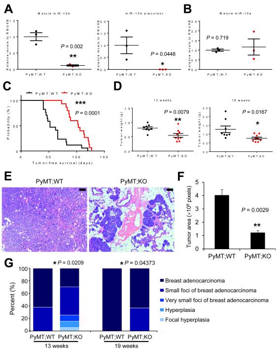
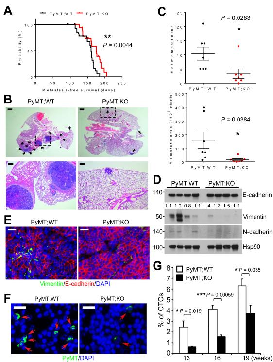
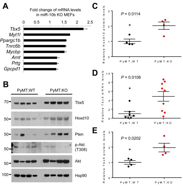
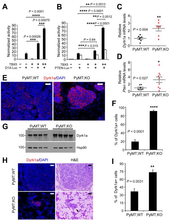
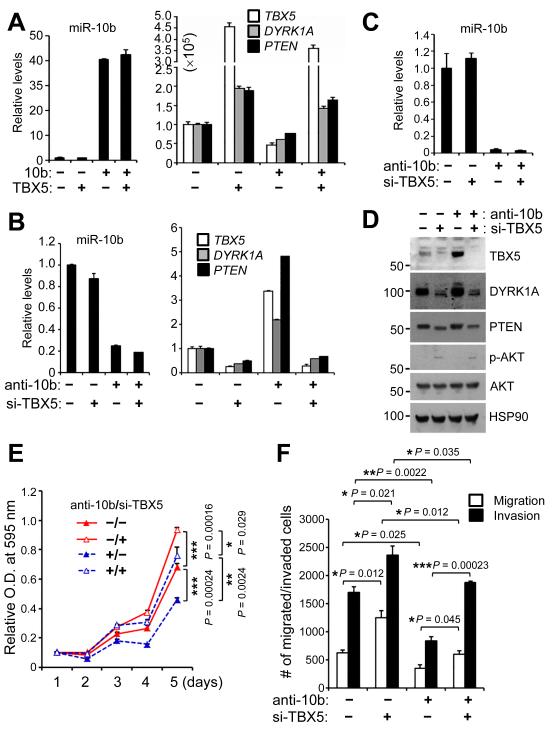
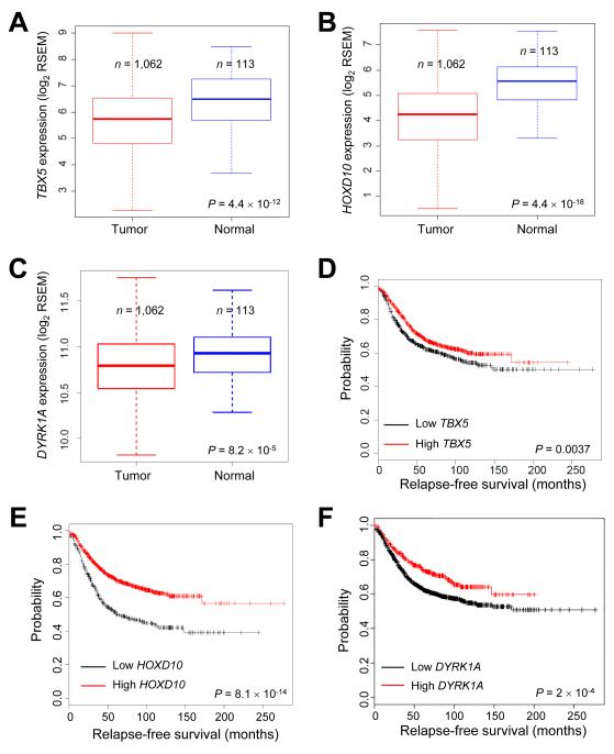
The invasive and metastatic properties of many human tumors have been associated with upregulation of the miRNA miR-10b, but its functional contributions in this setting have not been fully unraveled. Here, we report the generation of miR-10b-deficient mice, in which miR-10b is shown to be largely dispensable for normal development but critical to tumorigenesis. Loss of miR-10b delays oncogene-induced mammary tumorigenesis and suppresses epithelial-mesenchymal transition, intravasation, and metastasis in a mouse model of metastatic breast cancer. Among the target genes of miR-10b, the tumor suppressor genes Tbx5 and Pten and the metastasis suppressor gene Hoxd10 are significantly upregulated by miR-10b deletion. Mechanistically, miR-10b promotes breast cancer cell proliferation, migration, and invasion through inhibition of the expression of the transcription factor TBX5, leading to repression of the tumor suppressor genes DYRK1A and PTEN In clinical specimens of breast cancer, the expression of TBX5, HOXD10, and DYRK1A correlates with relapse-free survival and overall survival outcomes in patients. Our results establish miR-10b as an oncomiR that drives metastasis, termed a metastamiR, and define the set of critical tumor suppressor mechanisms it overcomes to drive breast cancer progression. Cancer Res; 76(21); 6424-35. ©2016 AACR.


Figures

Figure 1
Figure 1. Targeted deletion of miR-10b in mice
(A) The targeting strategy used to generate miR-10b knockout alleles. (B) PCR-based genotyping of wild-type (+), floxed (f), and deleted (-) alleles of miR-10b. (C) Northern blot analysis demonstrates miR-10b deletion in the kidney, testis, ovary, and embryo of age- and sex-matched wild-type (WT) and miR-10b−/− (KO) mice. (D) Spleen weight (relative to body weight) of age- and sex-matched wild-type (WT) and miR-10b−/− (KO) mice. n = 8 mice per group. (E) Gross examination (top) and H&E staining (bottom) of spleens of wild-type (WT) and miR-10b−/− (KO) mice. Scale bars, 200 μm. (F) Germinal center areas were quantitated by pixel counts. n ≥ 4 mice per group. P values in (D) and (F) were from a two-tailed, unpaired t-test.
Figure 2
Figure 2. Deletion of miR-10b impedes mammary tumor initiation, growth, and progression
(A, B) qPCR of mature miR-10b (A, left panel), the miR-10b precursor (A, right panel), and mature miR-10a (B) in mammary tumors of age-matched MMTV-PyMT;miR-10b WT and MMTV-PyMT;miR-10b KO mice. n = 3 mice per group. (C) Mammary tumor-free survival of MMTV-PyMT;miR-10b WT (n = 17) and MMTV-PyMT;miR-10b KO (n = 15) mice. The P value was from a log-rank test. (D) Mammary tumor weight of MMTV-PyMT;miR-10b WT and MMTV-PyMT;miR-10b KO mice at 13 and 16 weeks of age. n ≥ 8 mice per group. (E, F) H&E staining (E) and tumor area quantification (F) of mammary tissue sections from 13-week-old MMTV-PyMT;miR-10b WT and MMTV-PyMT;miR-10b KO mice. Scale bars in (E), 100 μm. n = 3 mice per group in (F). (G) Histopathological examination of mammary glands (n = 16, 20, 10, and 11 mice per group, from left to right). P values were from Wilcoxon rank-sum test. P values in (A), (B), (D), and (F) were from a two-tailed, unpaired t-test.
Figure 3
Figure 3. Deletion of miR-10b inhibits EMT, intravasation, and metastasis
(A) Metastasis-free survival of MMTV-PyMT;miR-10b WT (n = 67) and MMTV-PyMT;miR-10b KO (n = 53) mice. The P value was from a log-rank test. (B) H&E staining of lungs of MMTV-PyMT;miR-10b WT and MMTV-PyMT;miR-10b KO mice at 24-26 weeks of age. The bottom panels are high-magnification images of the boxed areas in the top panels. Arrows indicate metastatic foci. Scale bars, 1 mm (top) and 200 μm (bottom). (C) Quantitation of the number (top) and area (bottom) of metastatic foci in the lungs of MMTV-PyMT;miR-10b WT and MMTV-PyMT;miR-10b KO mice at 24-26 weeks of age. n = 7 mice per group. (D, E) Immunoblotting (D) and immunofluorescent staining (E) of EMT markers in mammary tumors of MMTV-PyMT;miR-10b WT and MMTV-PyMT;miR-10b KO mice. Scale bars in (E), 200 μm. (F, G) Circulating tumor cells (CTCs) were immunostained with a PyMT-specific antibody (green) and nuclei were stained with DAPI (blue) (F). The percentages of CTCs were quantitated (G). Scale bars in (F), 20 μm. n ≥ 4 mice per group in (G). P values in (C) and (G) were from a two-tailed, unpaired t-test.
Figure 4
Figure 4. Tbx5, Hoxd10, and Pten are upregulated by miR-10b deletion in mice
(A) qPCR of miR-10b targets in miR-10b-null MEFs. (B) Immunoblotting of Tbx5, Hoxd10, Pten, phospho-Akt, Akt, and Hsp90 in mammary tumors of MMTV-PyMT;miR-10b WT and MMTV-PyMT;miR-10b KO mice at 24-26 weeks of age. n = 4 mice per group. (C) Quantification of Hoxd10 protein levels in (B). (D) qPCR of Tbx5 in mammary tumors of MMTV-PyMT;miR-10b WT and MMTV-PyMT;miR-10b KO mice at 24-26 weeks of age. n ≥ 7 mice per group. (E) Quantification of Tbx5 protein levels in (B). P values in (C) – (E) were from a two-tailed, unpaired t-test.
Figure 5
Figure 5. DYRK1A and PTEN are TBX5 target genes and are upregulated in mammary tumors and lung metastases of MMTV-PyMT;miR-10b−/− mice
(A, B) Luciferase reporter assays show that the promoters of DYRK1A (A) and PTEN (B) are activated by TBX5 in a dose-dependent manner. D1A-Luc and PTEN-Luc are reporter constructs containing the human DYRK1A (1,497 bp) and PTEN (black bars, 1,064 bp; white bars, 1,978 bp) promoter regions, respectively. (C, D) qPCR of Dyrk1a (C) and Pten (D) in mammary tumors of MMTV-PyMT;miR-10b WT and MMTV-PyMT;miR-10b KO mice. (E, F) Immunofluorescent staining of Dyrk1a (E) and the percentages of Dyrk1a-positive cells (F) in mammary tumors of age-matched MMTV-PyMT;miR-10b WT and MMTV-PyMT;miR-10b KO mice. Scale bars in (E), 50 μm. n = 4 mice per group in (F). (G) Immunoblotting of Dyrk1a and Hsp90 in mammary tumors of MMTV-PyMT;miR-10b WT and MMTV-PyMT;miR-10b KO mice 24-26 weeks of age. n = 4 mice per group. (H, I) Immunofluorescent staining of Dyrk1a (H) and the percentages of Dyrk1a-positive cells (I) in lung metastases of age-matched MMTV-PyMT;miR-10b WT and MMTV-PyMT;miR-10b KO mice. Scale bars in (H), 50 μm. n = 4 mice per group in (I). P values in (A) – (D), (F), and (I) were from a two-tailed, unpaired t-test.
Figure 6
Figure 6. TBX5 is a functional target of miR-10b
(A) qPCR of miR-10b, TBX5, DYRK1A, and PTEN in 293FT cells transfected with miR-10b and TBX5, alone or in combination. (B) qPCR of miR-10b, TBX5, DYRK1A, and PTEN in 293FT cells transfected with miR-10b antisense inhibitors and TBX5 siRNA, alone or in combination. (C) qPCR of miR-10b in LM2 cells transfected with miR-10b antisense inhibitors and TBX5 siRNA, alone or in combination. (D) Immunoblotting of TBX5, DYRK1A, PTEN, phospho-AKT, AKT, and HSP90 in LM2 cells transfected with miR-10b antisense inhibitors and TBX5 siRNA, alone or in combination. (E, F) Growth curves (E) and migration and invasion assays (F) of LM2 cells transfected with miR-10b antisense inhibitors and TBX5 siRNA, alone or in combination. n = 4 and 3 wells per group in (E) and (F), respectively. P values were from a two-tailed, unpaired t-test.
Figure 7
Figure 7. TBX5, HOXD10, and DYRK1A are downregulated in human breast cancer and are associated with relapse-free survival
(A-C) Box plots comparing TBX5 (A), HOXD10 (B), or DYRK1A (C) expression in normal breast tissues and breast tumors, based on the RNA-Seq data from TCGA. The boxes show the median and the interquartile range. The whiskers show the minimum and maximum. P values were from a two-tailed, unpaired t-test. (D-F) Kaplan-Meier curves of relapse-free survival of breast cancer patients stratified by TBX5 (D), HOXD10 (E), or DYRK1A (F) expression levels. n = 1,660 patients. P values were from a log-rank test.

References

    1. Bartel DP. MicroRNAs: genomics, biogenesis, mechanism, and function. Cell. 2004;116:281–97. - PubMed
    1. Ma L, Weinberg RA. Micromanagers of malignancy: role of microRNAs in regulating metastasis. Trends Genet. 2008;24:448–56. - PubMed
    1. Piao HL, Ma L. Non-coding RNAs as regulators of mammary development and breast cancer. J Mammary Gland Biol Neoplasia. 2012;17:33–42. - PMC - PubMed
    1. Zhang J, Ma L. MicroRNA control of epithelial-mesenchymal transition and metastasis. Cancer Metastasis Rev. 2012;31:653–62. - PMC - PubMed
    1. Ma L, Teruya-Feldstein J, Weinberg RA. Tumour invasion and metastasis initiated by microRNA-10b in breast cancer. Nature. 2007;449:682–8. - PubMed

MeSH terms