Osteonecrosis of the Jaw in the Absence of Antiresorptive or Antiangiogenic Exposure: A Series of 6 Cases
- PMID: 27569557
- PMCID: PMC5527276
- DOI: 10.1016/j.joms.2016.07.019
Osteonecrosis of the Jaw in the Absence of Antiresorptive or Antiangiogenic Exposure: A Series of 6 Cases
Abstract
Purpose: Medication-related osteonecrosis of the jaws (MRONJ) is a well-described complication of antiresorptive and antiangiogenic medications. Although osteonecrosis can be associated with other inciting events and medications, such as trauma, infection, steroids, chemotherapy, and coagulation disorders, these are rarely reported in the literature.
Materials and methods: This is a six case series of MRONJ associated with medications other than antiresorptive or antiangiogenic drugs.
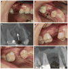
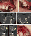
Results: Patient demographics, inciting event, location, stage, imaging findings, and outcome are reported.
Conclusion: With the continued development and clinical use of new biologic medications for diseases such as cancer and rheumatoid arthritis, it is important to continue to evaluate their effects on the oral cavity. The degree of risk for osteonecrosis in patients taking these new classes of drugs is uncertain but warrants awareness and monitoring.
Copyright © 2016 American Association of Oral and Maxillofacial Surgeons. Published by Elsevier Inc. All rights reserved.
Figures


Similar articles
-
Diagnosis and Staging of Medication-Related Osteonecrosis of the Jaw.Oral Maxillofac Surg Clin North Am. 2015 Nov;27(4):479-87. doi: 10.1016/j.coms.2015.06.008. Epub 2015 Aug 18. Oral Maxillofac Surg Clin North Am. 2015. PMID: 26293329 Review.
-
Methotrexate-associated osteonecrosis of the jaw: A report of two cases.Oral Surg Oral Med Oral Pathol Oral Radiol. 2017 Dec;124(6):e283-e287. doi: 10.1016/j.oooo.2017.09.005. Epub 2017 Sep 21. Oral Surg Oral Med Oral Pathol Oral Radiol. 2017. PMID: 29056286
-
Osteonecrosis of jaw beyond antiresorptive (bone-targeted) agents: new horizons in oncology.Expert Opin Drug Saf. 2016 Jul;15(7):925-35. doi: 10.1080/14740338.2016.1177021. Epub 2016 May 3. Expert Opin Drug Saf. 2016. PMID: 27074901 Review.
-
The Frequency of Medication-related Osteonecrosis of the Jaw and its Associated Risk Factors.Oral Maxillofac Surg Clin North Am. 2015 Nov;27(4):509-16. doi: 10.1016/j.coms.2015.06.003. Epub 2015 Sep 9. Oral Maxillofac Surg Clin North Am. 2015. PMID: 26362367 Review.
-
Radiographic Evaluation of Medication-Related Osteonecrosis of the Jaw (MRONJ) With Different Primary Cancers and Medication Therapies.Cureus. 2023 Aug 1;15(8):e42830. doi: 10.7759/cureus.42830. eCollection 2023 Aug. Cureus. 2023. PMID: 37664344 Free PMC article.
Cited by
-
Are medication-induced salivary changes the culprit of osteonecrosis of the jaw? A systematic review.Front Med (Lausanne). 2023 Aug 31;10:1164051. doi: 10.3389/fmed.2023.1164051. eCollection 2023. Front Med (Lausanne). 2023. PMID: 37720502 Free PMC article.
-
Management of dental care of patients on immunosuppressive drugs for chronic immune-related inflammatory diseases: a survey of French dentists' practices.BMC Oral Health. 2023 Aug 9;23(1):545. doi: 10.1186/s12903-023-03258-7. BMC Oral Health. 2023. PMID: 37559031 Free PMC article. Review.
-
Surgical and conservative treatment outcomes of medication-related osteonecrosis of the jaw located at tori: a retrospective study.Oral Maxillofac Surg. 2024 Sep;28(3):1117-1125. doi: 10.1007/s10006-024-01214-5. Epub 2024 Feb 29. Oral Maxillofac Surg. 2024. PMID: 38418702 Free PMC article.
-
Complications of invasive oral procedures in patients with immune-mediated inflammatory disorders treated with biological and conventional disease-modifying antirheumatic drugs or glucocorticoids: a scoping review of the literature.BMC Oral Health. 2025 Mar 27;25(1):442. doi: 10.1186/s12903-024-05414-z. BMC Oral Health. 2025. PMID: 40148875 Free PMC article.
-
Zoledronate treatment duration is linked to bisphosphonate-related osteonecrosis of the jaw prevalence in rice rats with generalized periodontitis.Oral Dis. 2019 May;25(4):1116-1135. doi: 10.1111/odi.13052. Epub 2019 Feb 19. Oral Dis. 2019. PMID: 30712276 Free PMC article.
References
-
- Henry DH, Costa L, Goldwasser F, et al. Randomized, double-blind study of denosumab versus zoledronic acid in the treatment of bone metastases in patients with advanced cancer (excluding breast and prostate cancer) or multiple myeloma. J Clin Oncol. 2011;29:1125. - PubMed
-
- Lipton A, Fizazi K, Stopeck AT, et al. Superiority of denosumab to zoledronic acid for prevention of skeletal-related events: A combined analysis of 3 pivotal, randomised, phase 3 trials. Eur J Cancer. 2012;48:3082. - PubMed
-
- Marx RE, Sawatari Y, Fortin M, et al. Bisphosphonate-induced exposed bone (osteonecrosis/osteopetrosis) of the jaws: Risk factors, recognition, prevention, and treatment. J Oral Maxillofac Surg. 2005;63:1567. - PubMed
-
- Stopeck AT, Lipton A, Body JJ, et al. Denosumab compared with zoledronic acid for the treatment of bone metastases in patients with advanced breast cancer: A randomized, double-blind study. J Clin Oncol. 2010;28:5132. - PubMed
Publication types
MeSH terms
Grants and funding
LinkOut - more resources
Full Text Sources
Other Literature Sources
Medical

