. 2016 Sep 14;20(3):296-306.
doi: 10.1016/j.chom.2016.07.018. Epub 2016 Aug 25.

The Yersinia Virulence Factor YopM Hijacks Host Kinases to Inhibit Type III Effector-Triggered Activation of the Pyrin Inflammasome

Affiliations

The Yersinia Virulence Factor YopM Hijacks Host Kinases to Inhibit Type III Effector-Triggered Activation of the Pyrin Inflammasome

Lawton K Chung et al. Cell Host Microbe. .

Abstract

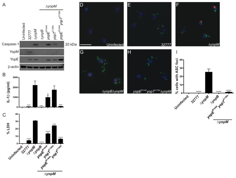
Pathogenic Yersinia, including Y. pestis, the agent of plague in humans, and Y. pseudotuberculosis, the related enteric pathogen, deliver virulence effectors into host cells via a prototypical type III secretion system to promote pathogenesis. These effectors, termed Yersinia outer proteins (Yops), modulate multiple host signaling responses. Studies in Y. pestis and Y. pseudotuberculosis have shown that YopM suppresses infection-induced inflammasome activation; however, the underlying molecular mechanism is largely unknown. Here we show that YopM specifically restricts the pyrin inflammasome, which is triggered by the RhoA-inactivating enzymatic activities of YopE and YopT, in Y. pseudotuberculosis-infected macrophages. The attenuation of a yopM mutant is fully reversed in pyrin knockout mice, demonstrating that YopM inhibits pyrin to promote virulence. Mechanistically, YopM recruits and activates the host kinases PRK1 and PRK2 to negatively regulate pyrin by phosphorylation. These results show how a virulence factor can hijack host kinases to inhibit effector-triggered pyrin inflammasome activation.


Figures

Figure 1
Figure 1. YopM inhibits activation of the pyrin inflammasome
LPS-primed wild-type or knock-out bone marrow-derived macrophages (BMDMs) were infected with the indicated Y. pseudotuberculosis strains for 90 min at a multiplicity of infection (MOI) of 30. (A,D) Caspase-1 processing in infected lysates was determined by Western blot (WB) analysis. (B,E) Secreted interleukin (IL)-1β was measured by ELISA and (C,F) cell-death was quantified by lactate dehydrogenase (LDH) release. Data in (B–C) and (E–F) represent average values ± SEM from three independent experiments compared to yopM mutant-infected BMDMs as analyzed by two- or one-way ANOVA, respectively. *, P < 0.05; ** P < 0.01; ****, P < 0.0001. See also Figure S1 and Figure S2.
Figure 2
Figure 2. YopM inhibits pyrin to counteract host protection to Yersinia
12–16 week old C57BL/6 or Mefv−/− mice were infected intravenously by tail-vein injection with approximately 2000 CFU of wild-type Y. pseudotuberculosis or a yopM mutant. (A–B) Time-to-death of infected mice was monitored for 21 days. Results are plotted to day 15 and pooled from two independent experiments with groups of four to five mice (n = 89). (C–D) Enumeration of bacterial burdens in the spleens and livers of infected mice at 5 days post-infection by CFU assays. Results are pooled from two independent experiments with groups of four mice (n = 8). Results in (A–B) were analyzed using the log-rank test while data in (C–D) were analyzed by Mann-Whitney test. *, P < 0.05; ***, P < 0.001; ****, P < 0.0001; ns, not significant.
Figure 3
Figure 3. The catalytic activities of YopE and YopT stimulate the pyrin inflammasome
LPS-primed BMDMs were infected with the indicated Y. pseudotuberculosis strains for 90 min at an MOI of 30. (A) Caspase-1 processing in infected lysates was determined by WB analysis. (B) Secreted IL-1β and (C) cytotoxicity was quantified by ELISA or LDH release, respectively. (D–H) Fluorescence imaging of Yersinia-infected BMDMs. ASC, red; Yersinia, green; nuceli, blue. Scale bar represents 10 μm. (I) Quantification of BMDMs containing ASC foci from images in (D–H). Data in (B–C) and (I) represent average values ± SEM from three independent experiments compared to yopM mutant-infected BMDMs as analyzed by one-way ANOVA. *, P < 0.05; *** P < 0.001; ****, P < 0.0001. See also Figure S3.
Figure 4
Figure 4. The catalytic activities of YopE and YopT drive responses that attenuate a Yersinia yopM mutant in vivo
C57BL/6 mice were infected intravenously via tail vein injection with ~5,000 CFU of the indicated Y. pseudotuberculosis strains and time-to-death was monitored for 21 days. Results are plotted to day 15 and pooled from two independent experiments with four to six mice per group (n = 810). Survival curves were analyzed using the log-rank test. ****, P < 0.0001. See also Figure S4.
Figure 5
Figure 5. YopM associates with host protein kinases and hijacks PRK to negatively regulate pyrin by phosphorylation
(A) WB analysis was performed to detect host proteins that bound to GST or GST-YopM32777 in lysates (starting material) from HEK293T cells transfected to express Myc-tagged murine pyrin. (B) In vitro kinase assays were performed with proteins bound to GST or GST-YopM32777 in lysates of HEK293T cells that were transfected to express Myc-pyrin. Reactions performed in the presence of [γ-32P]ATP were directly loaded or immunoprecipitated using an anti-Myc antibody prior to analysis by SDS-PAGE and autoradiography. Parallel kinase reactions were performed using cold ATP and analyzed by WB analysis to verify presence of PRK2, pyrin (Myc), RSK1 and YopM. Arrowhead indicates phosphorylated Myc-pyrin. (C) LPS-primed BMDMs were infected with the indicated Y. pseudotuberculosis strains for 90 min at an MOI of 30 in the presence or absence of PRK (PKC412) or RSK (BI-D1870) inhibitors and caspase-1 processing in infected lysates was detected by WB analysis. (D) WB analysis of IL-1β in the supernatants and lysates of Yersinia-infected LPS-primed BMDMs that were transiently transfected with control siRNAs or siRNAs targeting PRK1 and PRK2 prior to infection. See also Figure S5.
Figure 6
Figure 6. YopM increases PRK-mediated pyrin phosphorylation
Recombinant Myc-tagged N-terminal pyrin (amino acids 1–330) was incubated with either purified PRK1 or PRK2 and recombinant GST or GST-YopM32777, after which phosphorylation of pyrin was assessed by WB analysis for phosphorylated serine.

Comment in

References

    1. AKTORIES K. Bacterial protein toxins that modify host regulatory GTPases. Nat Rev Microbiol. 2011;9:487–98. - PubMed
    1. AKULA MK, SHI M, JIANG Z, FOSTER CE, MIAO D, LI AS, ZHANG X, GAVIN RM, FORDE SD, GERMAIN G, CARPENTER S, ROSADINI CV, GRITSMAN K, CHAE JJ, HAMPTON R, SILVERMAN N, GRAVALLESE EM, KAGAN JC, FITZGERALD KA, KASTNER DL, GOLENBOCK DT, BERGO MO, WANG D. Control of the innate immune response by the mevalonate pathway. Nat Immunol 2016 - PMC - PubMed
    1. AUBERT DF, XU H, YANG J, SHI X, GAO W, LI L, BISARO F, CHEN S, VALVANO MA, SHAO F. A Burkholderia Type VI Effector Deamidates Rho GTPases to Activate the Pyrin Inflammasome and Trigger Inflammation. Cell Host Microbe 2016 - PubMed
    1. BERGSBAKEN T, FINK SL, COOKSON BT. Pyroptosis: host cell death and inflammation. Nat Rev Microbiol. 2009;7:99–109. - PMC - PubMed
    1. BERNEKING L, SCHNAPP M, RUMM A, TRASAK C, RUCKDESCHEL K, ALAWI M, GRUNDHOFF A, KIKHNEY AG, KOCH-NOLTE F, BUCK F, PERBANDT M, BETZEL C, SVERGUN DI, HENTSCHKE M, AEPFELBACHER M. Immunosuppressive Yersinia Effector YopM Binds DEAD Box Helicase DDX3 to Control Ribosomal S6 Kinase in the Nucleus of Host Cells. PLoS Pathog. 2016;12:e1005660. - PMC - PubMed

MeSH terms