. 2018 Jan;15(1):38-47.
doi: 10.1038/cmi.2016.43. Epub 2016 Aug 29.

Cold-inducible RNA-binding protein activates splenic T cells during sepsis in a TLR4-dependent manner

Affiliations

Cold-inducible RNA-binding protein activates splenic T cells during sepsis in a TLR4-dependent manner

Alexandra C Bolognese et al. Cell Mol Immunol. 2018 Jan.

Abstract

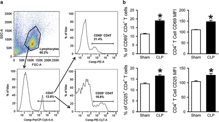
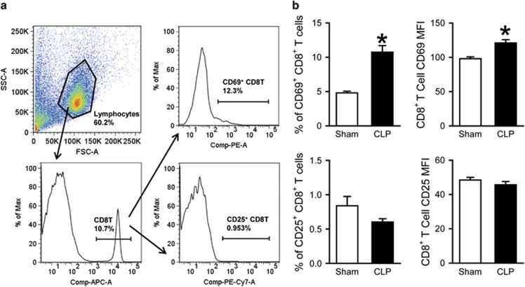
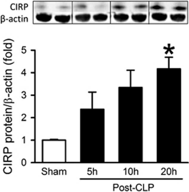
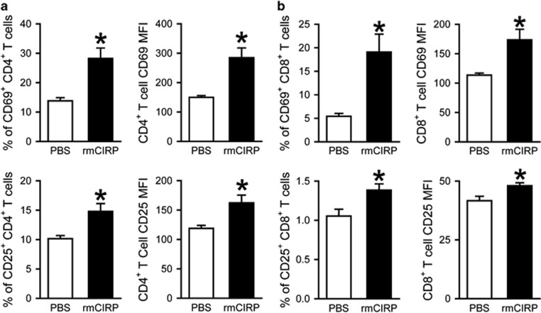
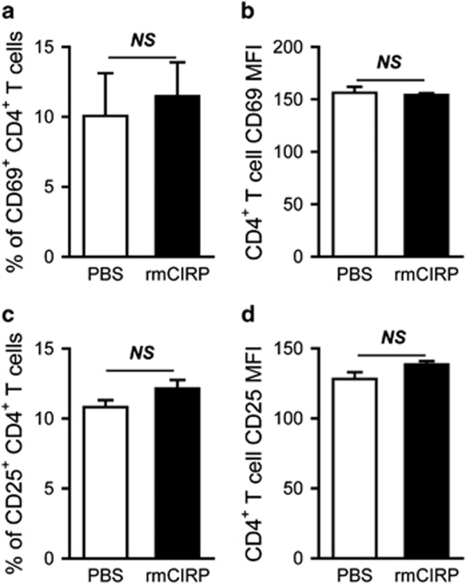
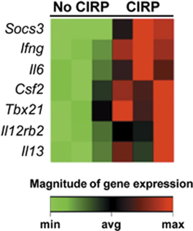
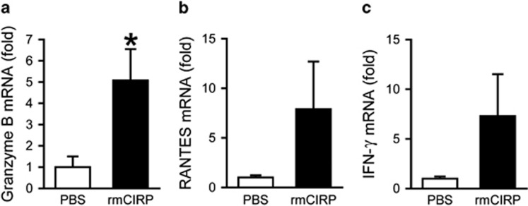
Cold-inducible RNA-binding protein (CIRP) is a novel inflammatory mediator that stimulates the release of proinflammatory cytokines from macrophages in sepsis. Given the immune dysregulation that characterizes sepsis, the effect of CIRP on other immune cells is an area of increasing interest that has not yet been studied. In the present study, we hypothesized that extracellular CIRP promotes activation of T lymphocytes in the spleen during sepsis. We observed that mice subjected to sepsis by cecal ligation and puncture showed significantly higher expression of the early activation markers CD69 and CD25 at 20 h on CD4+ splenic T cells, and significantly higher CD69 expression on CD8+ splenic T cells compared with sham-operated controls. Furthermore, at 20 h after receiving intravenous injection of recombinant murine CIRP (rmCIRP, 5 mg/kg body weight (BW)) or PBS (vehicle), those mice receiving rmCIRP showed significantly increased expression of CD69 and CD25 on both CD4+ and CD8+ splenic T cells. This effect, however, was not seen in TLR4-deficient mice after rmCIRP injection. In addition, treatment with CIRP predisposed CD4+ T cells to a Th1 hyperinflammatory response profile, and influenced CD8+ T cells toward a cytotoxic profile. Taken together, our findings indicate that CIRP is a proinflammatory mediator that plays an important role in T-cell dysregulation during sepsis in a TLR4-dependent manner.


Conflict of interest statement

The authors declare no conflict of interest.

Figures

Figure 1
Figure 1
Activation of CD4+ splenic T cells after CLP. Mice underwent CLP or sham operation; spleens were collected 20 h later and processed to isolate lymphocytes. (a) A representative sample showing flow cytometry gating strategy. Total splenocytes were gated for the live lymphocyte population based on FSC and SSC characteristics. Lymphocytes were subsequently gated for the PerCP/Cy5.5-CD4-positive cell population and further gated into those staining positively for PE-CD69 and PE/Cy7-CD25. (b) Flow cytometric analysis of surface CD69 and CD25 expression on the gated splenic CD4+ T cells. Percentages of CD69+ and CD25+ cells and mean fluorescence intensity (MFI) of CD69 and CD25 expression are shown. Data expressed as mean±s.e.m. (n=5 per group) and compared by Student’s t-test. *P<0.05 vs Sham. CLP, cecal ligation and puncture; FSC, forward scatter; SSC, side scatter.
Figure 2
Figure 2
Activation of CD8+ splenic T cells after CLP. Mice underwent CLP or sham operation; spleens were collected 20 h later and processed to isolate lymphocytes. (a) A representative sample showing flow cytometry gating strategy. Total splenocytes were gated for the live lymphocyte population based on FSC and SSC characteristics. Lymphocytes were subsequently gated for the APC-CD8-positive cell population and further gated into those staining positively for PE-CD69 and PE/Cy7-CD25. (b) Flow cytometric analysis of surface CD69 and CD25 expression on the gated splenic CD8+ T cells. Percentages of CD69+ and CD25+ cells and MFI of CD69 and CD25 expression are shown. Data expressed as mean±s.e.m. (n=5 per group) and compared by Student’s t-test. *P<0.05 vs Sham. CLP, cecal ligation and puncture; FSC, forward scatter; SSC, side scatter.
Figure 3
Figure 3
Protein levels of CIRP in the spleen after CLP. Mice underwent CLP or sham operation; spleens were collected at 5, 10 and 20 h after CLP and CIRP protein levels were determined by western blot. A representative blot is shown along with a graph representing densitometry analysis. Data expressed as mean±s.e.m. (n=2 mice in sham group and 3 mice per CLP group). *P<0.05 vs Sham by Student’s t-test. CLP, cecal ligation and puncture; CIRP, cold-inducible RNA-binding protein.
Figure 4
Figure 4
Activation of CD4+ and CD8+ splenic T cells by rmCIRP. rmCIRP (5 mg/kg BW) or PBS was intravenously injected in mice; spleens were collected 20 h later and processed to isolate lymphocytes. Flow cytometric analysis of surface CD69 and CD25 expression on the gated splenic CD4+ T cells (a) and CD8+ T cells (b) from rmCIRP- and PBS-injected mice. Percentages of CD69+ and CD25+ cells and mean fluorescence intensity (MFI) of CD69 and CD25 expression are shown. Data expressed as mean±s.e.m. (n=4–5 per group) and compared by Student’s t-test. *P<0.05 vs PBS. BW, body weight; rmCIRP, recombinant murine CIRP; PBS, phosphate-buffered solution.
Figure 5
Figure 5
Mediation of splenic CD4+ T-cell activation by TLR4. rmCIRP (5 mg/kg BW) or PBS was intravenously injected in Tlr4−/− mice; spleens were collected 20 h later and processed to isolate lymphocytes. Flow cytometric analysis of surface CD69 and CD25 expression on the gated splenic CD4+ T cells from PBS- and rmCIRP-injected Tlr4−/− mice. Percentages of CD69+ (a) and CD25+ (c) cells and MFI of CD69 (b) and CD25 (d) expression on gated CD4+ T cells are shown. Data expressed as mean±s.e.m. (n=4–5 per group) and compared by Student’s t-test. BW, body weight; rmCIRP, recombinant murine CIRP; PBS, phosphate-buffered solution; MFI, mean fluorescence intensity; NS, no significance.
Figure 6
Figure 6
Clustergram of genes upregulated in CD4+ T cells after treatment with rmCIRP. Purified splenic CD4+ T cells (1 × 10/ml/well) were treated with rmCIRP (1 μg/ml) or PBS for 20 h during plate-bound anti-CD3/CD28 (1 μg/ml each) stimulation. An 84-gene PCR array was used to examine Th1 vs Th2 response profiles for CD4+ T cells. A clustergram featuring the seven genes that were significantly upregulated is shown (n=3 per group, P<0.05 by Student’s t-test). Of these, four are Th1-related genes (Ifng, Csf2, Tbx21 and Il12rb2), two are CD4 T-cell markers (Socs3 and Il6), and one is a Th2-related cytokine (Il13). No genes were downregulated. rmCIRP, recombinant murine CIRP; PCR, polymerase chain reaction.
Figure 7
Figure 7
Increased gene expression in CD8+ T cells after treatment with rmCIRP. Purified splenic CD8+ T cells (1 × 10⁶/ml/well) were treated with rmCIRP (1 μg/ml) or PBS for 20 h during plate-bound anti-CD3/CD28 (1 μg/ml each) stimulation. Fold increase in expression of granzyme B (a), RANTES (b) and IFN-γ (c) mRNA is shown relative to cells treated with PBS. β-actin was used as the reference gene. Data expressed as mean±s.e.m. (n=4 per group) and compared by Student’s t-test. *P<0.05 vs PBS. rmCIRP, recombinant murine CIRP; PBS, phosphate-buffered solution.

References

    1. Singer M, Deutschman CS, Seymour CW, Shankar-Hari M, Annane D, Bauer M et al. The third international consensus definitions for sepsis and septic shock (Sepsis-3). JAMA 2016; 315: 801–810. - PMC - PubMed
    1. Angus DC, van der Poll T. Severe sepsis and septic shock. N Engl J Med 2013; 369: 2063. - PubMed
    1. Fleischmann C, Scherag A, Adhikari NK, Hartog CS, Tsaganos T, Schlattmann P et al. Assessment of global incidence and mortality of hospital-treated sepsis. Current estimates and limitations. Am J Respir Crit Care Med 2016; 193: 259–272. - PubMed
    1. Scumpia PO, Delano MJ, Kelly KM, O'Malley KA, Efron PA, McAuliffe PF et al. Increased natural CD4+CD25+ regulatory T cells and their suppressor activity do not contribute to mortality in murine polymicrobial sepsis. J Immunol. 2006; 177: 7943–7949. - PubMed
    1. Martin GS. Sepsis, severe sepsis and septic shock: changes in incidence, pathogens and outcomes. Expert Rev Anti Infect Ther 2012; 10: 701–706. - PMC - PubMed

Publication types

MeSH terms