. 2016 Sep 8;166(6):1485-1499.e15.
doi: 10.1016/j.cell.2016.07.046. Epub 2016 Aug 25.

γδ T Cells Support Pancreatic Oncogenesis by Restraining αβ T Cell Activation

Affiliations

γδ T Cells Support Pancreatic Oncogenesis by Restraining αβ T Cell Activation

Donnele Daley et al. Cell. .

Erratum in

  • γδ T Cells Support Pancreatic Oncogenesis by Restraining αβ T Cell Activation.
    Daley D, Zambirinis CP, Seifert L, Akkad N, Mohan N, Werba G, Barilla R, Torres-Hernandez A, Hundeyin M, Kumar Mani VR, Avanzi A, Tippens D, Narayanan R, Jang JE, Newman E, Pillarisetty VG, Dustin ML, Bar-Sagi D, Hajdu C, Miller G. Daley D, et al. Cell. 2020 Nov 12;183(4):1134-1136. doi: 10.1016/j.cell.2020.10.041. Cell. 2020. PMID: 33186522 No abstract available.

Abstract

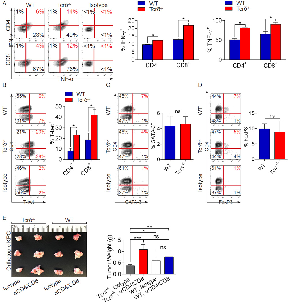
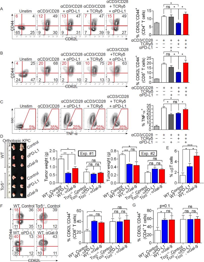
Inflammation is paramount in pancreatic oncogenesis. We identified a uniquely activated γδT cell population, which constituted ∼40% of tumor-infiltrating T cells in human pancreatic ductal adenocarcinoma (PDA). Recruitment and activation of γδT cells was contingent on diverse chemokine signals. Deletion, depletion, or blockade of γδT cell recruitment was protective against PDA and resulted in increased infiltration, activation, and Th1 polarization of αβT cells. Although αβT cells were dispensable to outcome in PDA, they became indispensable mediators of tumor protection upon γδT cell ablation. PDA-infiltrating γδT cells expressed high levels of exhaustion ligands and thereby negated adaptive anti-tumor immunity. Blockade of PD-L1 in γδT cells enhanced CD4(+) and CD8(+) T cell infiltration and immunogenicity and induced tumor protection suggesting that γδT cells are critical sources of immune-suppressive checkpoint ligands in PDA. We describe γδT cells as central regulators of effector T cell activation in cancer via novel cross-talk.

Keywords: Kras; cancer; checkpoint ligands.


Figures

Figure 1
Figure 1. γδT cells are ubiquitous and activated in human PDA
(a) Frozen sections of human PDA and normal pancreas were stained using a mAb specific for TCRγ/δ or isotype control. Representative images and quantitative data are shown. (b) Single cell suspensions from human PDA tumors and PBMC were co-stained for CD45, CD3, and TCRγ/δ. The percentage of γδT cells among CD3+ cells was calculated. Representative contour plots and summary data are shown. Each dot represents a different patient sample. (c) The percentage of PDA-infiltrating γδT cells among CD45+ cells was compared with tumor-infiltrating cells expressing select myeloid differentiation markers. (d) The percentage of PDA-infiltrating and PBMC γδT cells among CD3+ cells was compared with that of CD4+ and CD8+ αβT cell subsets in each respective compartment. (e) PBMC and PDA-infiltrating CD3+TCRγ/δ+ cells from PDA patients were gated and co-stained using mAbs specific for CD45RA and CD27. The gating paradigms for Tnaive, TCM, TEM, and TEM-RA populations are shown. Representative contour plots and quantitative data indicating the fraction of TEM γδT cells in each compartment are indicated. (f) PDA-infiltrating and PBMC γδT cells from PDA patients were stained using mAbs specific for CD62L and (g) Vγ9. Representative histograms and quantitative data are shown. Human data are based on tumor tissue or PBMC analyzed from 9–13 PDA patients (*p<0.05, **p<0.01, ***p<0.001).
Figure 2
Figure 2. γδT cells are highly prevalent and exhibit a uniquely activated phenotype in murine invasive PDA
(a) C57BL/6-Trdctm1Mal mice whose γδT cells express GFP were orthotopically implanted with KPC-derived tumor and imaged by intra-vital two-photon laser-scanning microscopy at 21 days. (b) WT mice were orthotopically implanted with KPC-derived tumor cells. On day 21, single cell suspensions of digested PDA tumors and splenocytes were co-stained for CD45, CD3, TCRγ/δ, CD4, and CD8 and analyzed by flow cytometry. Representative contour plots and quantitative data are shown. (c) WT mice were orthotopically implanted with KPC-derived tumor cells. On day 21 spleen (blue histograms) and PDA-infiltrating (red histograms) γδT cells were gated and tested for co-expression of select surface activation markers and Vγ chains. Representative histogram overlays and summary data from 5 mice are shown. (d) Spleen and PDA-infiltrating γδT cells from the same mice were tested for expression of IL-10, (e) IL-17, (f) NKG2D, (g) TLR4, TLR7, TLR9, and (h) CCR2, CCR5, and CCR6. Each experiment was repeated at least 3 times using 3–5 mice per data point (*p<0.05, **p<0.01).
Figure 3
Figure 3. Ablation of γδT cells protects against pancreatic oncogenesis in a slowly progressive model of PDA
(a) KC;Tcrδ+/+ and KC;Tcrδ−/− mice were sacrificed at 3, 6, or 9 months of life (n=10–12 mice/cohort). Representative H&E-stained frozen sections are shown. The percentage of pancreatic area occupied by intact acinar structures, and the fractions of ductal structures exhibiting normal morphology, ADM, or graded PanIN I-III lesions were calculated. (b) Weights of pancreata were compared in 3 month-old KC;Tcrδ+/+ and KC;Tcrδ−/− mice. (c) Pancreata from 9 month-old KC;Tcrδ+/+ and KC;Tcrδ−/− mice were assayed for peri-tumoral fibrosis using trichrome staining. (d) Kaplan-Meier survival analysis was performed for KC;Tcrδ+/+ (n=29) and KC;Tcrδ−/− (n=44) mice (p<0.0001). (e, f) KC;Tcrδ+/+ mice were treated with UC3-10A6 or isotype control for 8 weeks beginning at 6 weeks of life. (e) Representative H&E stained pancreatic sections are shown. The percentage of pancreatic area occupied by intact acinar structures, and the fractions of ductal structures exhibiting normal morphology, ADM, or graded PanIN I-III lesions were calculated. (f) Tumor weight was recorded (n=5/group; *p<0.05, **p<0.01).
Figure 4
Figure 4. γδT cell deletion results in massive CD4+ and CD8+ T cell infiltration and activation in invasive PDA
(a, b) WT and Tcrδ−/− mice were implanted with KPC-derived tumor cells. On day 21 mice were sacrificed. Frozen pancreatic sections were tested for (a) CD8+ and (b) CD4+ T cell infiltration by IHC (n=5/group). (c) CD8+ T cells infiltrating orthotopically-implanted KPC-derived tumors in WT and Tcrδ−/− mice were tested for expression of CD44, (d) ICOS, (e) CTLA-4, and (f) Granzyme B. (g) Similarly, CD4+ T cells infiltrating orthotopically-implanted KPC tumors in WT and Tcrδ−/− mice were tested for expression of CD44, (h) OX40, (i) PD-1, and (j) CD62L. Experiments were repeated more than 3 times with similar results using 5 mice per group (*p<0.05, **p<0.01, ***p<0.001).
Figure 5
Figure 5. γδT cell deletion results in CD4+ T cell Th1 differentiation, CD8+ T cell activation, and αβT cell-dependent tumor protection in invasive PDA
(a–d) WT and Tcrδ−/− mice were orthotopically implanted with KPC-derived tumor cells. On day 21, tumor-infiltrating CD4+ and CD8+ T cells were interrogated for (a) co-expression of TNF-α and IFN-γ, (b) expression of T-bet, (c) GATA-3, and (d) FoxP3. Representative contour plots and quantitative data are shown. Experiments were repeated twice with similar results (n=5/group; *p<0.05). (e) WT and Tcrδ−/− pancreata were orthotopically implanted with KPC-derived tumor cells and serially treated with α-CD4 and α-CD8 neutralizing mAbs or isotype controls. Pancreatic tumors were harvested at 3 weeks. Representative images and tumor weights are shown (n=5/group; *p<0.05, **p<0.01, ***p<0.001).
Figure 6
Figure 6. PDA-associated γδT cells express high levels of T cell exhaustion ligands in multiple murine tumor models and in human disease
(a) Expression of PD-L1 and (b) Galectin-9 were compared in pancreas and spleen γδT cells of 3-month-old KC mice by flow cytometry. Representative contour plots and quantitative data are shown (n=5/group). (c) WT mice were orthotopically implanted with KPC-derived tumor cells. Expression of PD-L1 and Galectin-9 were compared in PDA tumor cells, TAMs (Mφ), MDSC, and γδT cells on day 21 (n=5/group). (d) WT mice were orthotopically implanted with KPC-derived tumor cells. On day 21, spleen and PDA-infiltrating γδT cells were tested for expression of select activating ligands. Representative histograms and quantitative data are shown (n=5/group). (e) Orthotopic PDA-bearing WT and Tcrδ−/− mice were tested for expression of PD-L1 in tumor cells, TAMs, and MDSC (n=5/group). (f) WT, CCR2−/−, CCR5−/−, and CCR6−/− mice were orthotopically implanted with KPC-derived PDA cells (n=5/group). Animals were sacrificed at 3 weeks, and the fraction of tumor-infiltrating γδT cells expressing PD-L1 and (g) Galectin-9 were determined by flow cytometry. (h, i) PBMC γδT cells from healthy volunteers and PDA patients, and PDA-infiltrating γδT cells and were tested for expression of (h) PD-L1 and (i) Galectin-9. Representative histograms and quantitative data are shown (n=11 patients; *p<0.05, **p<0.01, ***p<0.001).
Figure 7
Figure 7. Exhaustion ligand blockade reverses the direct suppressive effects of γδT cells on αβT cells and on pancreatic tumorigenesis
(a) Splenic CD4+ or (b) CD8+ T cells from untreated WT mice were either unstimulated, or stimulated with αCD3/αCD28 alone or in co-culture with PDA-infiltrating γδT cells (5:1 ratio). αPD-L1 (10µg/ml) was selectively added to each group. The fraction of CD62LCD44+ cells were determined at 72h by flow cytometry. Representative contour plots and quantitative data are shown. (c) Similarly, CD4+ and CD8+ T cell expression of TNF-α was measured. Experiments were performed in quadruplicate and repeated 3 times. (d) WT and Tcrδ−/− mice were orthotopically implanted with KPC-derived tumor cells and serially treated with αPD-L1 or αGalectin-9 neutralizing mAbs, or respective isotype controls. Pancreatic tumors were harvested at 3 weeks. Representative gross images are shown (Experiment #1) as are quantitative data on tumor weights from 2 separate experiments using different stocks of KPC-derived tumor cells (n=5/group for each experiment). (e–g) WT and Tcrδ−/− pancreata were again orthotopically implanted with KPC-derived tumor cells and serially treated with αPD-L1 or αGalectin-9 neutralizing mAbs or the respective isotype controls. Pancreatic tumors were harvested at 3 weeks. (e) The fraction of PDA-infiltrating αβT cells among CD45+ leukocytes, and (f) CD8+ and (g) CD4+ T cell adoption of an activated CD62LCD44+ phenotype, were determined by flow cytometry (n=5/group; *p<0.05, **p<0.01, ***p<0.01).

Comment in

  • Pancreatic cancer: γδ T cells support PDAC.
    Dickson I. Dickson I. Nat Rev Gastroenterol Hepatol. 2016 Oct;13(10):560. doi: 10.1038/nrgastro.2016.156. Epub 2016 Sep 14. Nat Rev Gastroenterol Hepatol. 2016. PMID: 27625194 No abstract available.

References

    1. Andren-Sandberg A, Dervenis C, Lowenfels B. Etiologic links between chronic pancreatitis and pancreatic cancer. Scand J Gastroenterol. 1997;32:97–103. - PubMed
    1. Bayne LJ, Beatty GL, Jhala N, Clark CE, Rhim AD, Stanger BZ, Vonderheide RH. Tumor-derived granulocyte-macrophage colony-stimulating factor regulates myeloid inflammation and T cell immunity in pancreatic cancer. Cancer cell. 2012;21:822–835. - PMC - PubMed
    1. Bedrosian AS, Nguyen AH, Hackman M, Connolly MK, Malhotra A, Ibrahim J, Cieza-Rubio NE, Henning JR, Barilla R, Rehman A, et al. Dendritic cells promote pancreatic viability in mice with acute pancreatitis. Gastroenterology. 2011;141:1915–1926. e1911–e1914. - PMC - PubMed
    1. Clark CE, Hingorani SR, Mick R, Combs C, Tuveson DA, Vonderheide RH. Dynamics of the immune reaction to pancreatic cancer from inception to invasion. Cancer research. 2007;67:9518–9527. - PubMed
    1. Coffelt SB, Kersten K, Doornebal CW, Weiden J, Vrijland K, Hau CS, Verstegen NJ, Ciampricotti M, Hawinkels LJ, Jonkers J, et al. IL-17-producing gammadelta T cells and neutrophils conspire to promote breast cancer metastasis. Nature. 2015 - PMC - PubMed

Publication types