. 2015 Feb 10:135:40-51.
doi: 10.1016/j.minpro.2015.01.003.

Microwave propagation and absorption and its thermo-mechanical consequences in heterogeneous rocks

Affiliations

Microwave propagation and absorption and its thermo-mechanical consequences in heterogeneous rocks

R Meisels et al. Int J Miner Process. .

Abstract

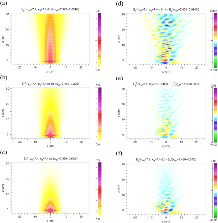
A numerical analysis in a two-component model rock is presented including the propagation and absorption of a microwave beam as well as the microwave-induced temperature and stress distributions in a consistent way. The analyses are two-dimensional and consider absorbing inclusions (discs) in a non-absorbing matrix representing the model of a heterogeneous rock. The microwave analysis (finite difference time domain - FDTD) is performed with values of the dielectric permittivity typical for hard rocks. Reflections at the discs/matrix interfaces and absorption in the discs lead to diffuse scattering with up to 20% changes of the intensity in the main beam compared to a homogeneous model rock. The subsequent thermo-mechanical finite element (FE) analysis indicates that the stresses become large enough to initiate damage. The results are supported by preliminary experiments on hard rock performed at 2.45 GHz.

Keywords: Microwave analysis; Microwave heating; Microwave irradiation experiments; Rock damage; Thermally induced stresses.


Figures

Fig. 1
Fig. 1
Two-dimensional model of a block of rock with statistical distribution of discs in a matrix (details see Fig. 2). The microwave source (not shown) is positioned at x = 0 and z = − 1 cm. Its width in x direction is 8.6 cm.
Fig. 2
Fig. 2
Enlarged section of the two-dimensional model rock. The grains in the matrix are represented by discs positioned on a square lattice with positional disorder. The crosses represent the lattice points, the dots the actual centers of the discs. δx and δz denote the deviation from the ideal position on a lattice point. After Meisels and Kuchar (2007).
Fig. 3
Fig. 3
Results for heterogeneity in the real parts of the permittivity and no loss (κm.i = κd,i = 0). (a) Distribution of Ey2 in the model rock and in front of it. κm = 5.4, κd = 9.4. Definition of Ey2 see Section 2. (b, c) Difference ΔEy2 of the distributions of Ey2 in the heterogeneous and homogeneous model. κm = 5.4, κd = 7.4 and 9.4, respectively. κeff calculated from Eq. (4). (d–f) Cuts along the z direction at x = 0 through the Ey2 and ΔEy2 distributions of (a), (b), and (c). In (d) the solid line is for the heterogeneous model, the dashed one for the homogeneous model.
Fig. 4
Fig. 4
Results for heterogeneity only due to absorption in the discs: κm,r = κd,r = 7.4, κd,i = 0.1, 0.88 and 2.0, κm,i = 0. (a–c) Distribution of Ey2 in the model rock and in front of it. (d–f) Difference between the distributions of Ey2 in the heterogeneous and homogeneous models. Note the change of the color scale.
Fig. 5
Fig. 5
Results for differences in the real as well as in the imaginary part of the permittivity of the matrix and the discs, i.e. heterogeneity due to reflection at the discs/matrix interfaces and absorption in the discs. (a–c) Distributions of Ey2 in the model rock and in front of it. (d–f) Distributions of ΔEy2. Permittivity values: κm,r = 7.1, κm,i = 0, and κeff,r = 7.4. Values of κeff,i (varied), and of κd,r and κd,i (calculated from the other values, Eq. (3)) are given in the figures.
Fig. 6
Fig. 6
Ey2 (a) and ΔEy2 (b) for a larger difference of the real parts (κm,r = 5.4, and κd,r = 9.4) than in Fig. 5; κd,i = 0.88. In this case κeff is calculated from the other values (Eq. (4)).
Fig. 7
Fig. 7
Pattern of the absorbed power density within the model rock. (a) Large penetration depth. (b) Small penetration depth. Compare Ey2 in Fig. 5(a) and (b). The color scale indicates the time-averaged absorbed power density in units of W/cm3 normalized to a time-averaged Ey2 of 1 V2/cm2 of the microwave source at x = 0 cm and z = − 1 cm. Note the difference in the values of the absorbed power. (c) Absorbed power density for a homogeneous reference case (cf. (b)).
Fig. 8
Fig. 8
Temperature distribution in °C after 15 s of microwave irradiation (17.5 kW) in the 2D model rock corresponding to Fig. 7(b). Sample size as in Fig. 3, Fig. 4, Fig. 5, Fig. 6, Fig. 7.
Fig. 9
Fig. 9
Maximum principal stresses in Pa in the model rock after 15 s of microwave irradiation calculated from the temperature distribution of Fig. 8. Dark red indicates values larger than 9 MPa.
Fig. 10
Fig. 10
Vector plot of the maximum principal stress (greater than tensile strength of 9 MPa) after 15 s of microwave irradiation in the magnified area shown in Fig. 9; the arrows indicate the direction of the normal to a potential crack plane.
Fig. 11
Fig. 11
Comparison of disc arrangement (circles) and the ΔEy2 distribution (color pattern). Enlarged section of Fig. 5(e) near the microwave source.
Fig. 12
Fig. 12
Comparison of density variations of the disc distribution (contour lines) and the ΔEy2 distribution as in Fig. 5(e) (color pattern). 0.34 is the average density (= filling factor). Permittivities of discs and matrix same as in Fig. 7. Near the boundary of the model rock (x = 0) the values of the density approach zero. Contours with values smaller than 0.3 are not shown here.
Fig. 13
Fig. 13
Spot on basalt irradiated with 2.45 GHz microwaves for 15 s. The power of the microwave source is 25 kW. Losses are estimated to be 30% yielding 17.5 kW available for absorption in the rock. Spallation is clearly visible. The irradiated area has approximately the size of the circle.
Fig. 14
Fig. 14
Three spots (indicated by red circles) on gabbro (a) and granite (b) after irradiation with 25 kW (power of the microwave source) for durations given in the figures. Cracks develop due to irradiation for more than 30 and 20 s, respectively, and extend far outside the irradiated area. Surface treated with penetration spray to provide better visibility of cracks.

References

    1. ABAQUS . Dassault Systèmes Simulia Corp; Providence, RI, USA: 2012. Abaqus v6.12 Documentation. (URL http://www.3ds.com/products-services/simulia/portfolio/abaqus/overview/)
    1. Ackermann R.J., Sorrell C.A. Thermal expansion and the high–low transformation in quartz. I. High-temperature x-ray studies. J. Appl. Crystallogr. 1974;7(5):461–467.
    1. Ali A.Y., Bradshaw S.M. Quantifying damage around grain boundaries in microwave treated ores. Chem. Eng. Process. Process Intensif. 2009;48(11–12):1566–1573.
    1. Ali A.Y., Bradshaw S.M. Bonded-particle modelling of microwave-induced damage in ore particles. Miner. Eng. 2010;23(10):780–790.
    1. Ali A.Y., Bradshaw S.M. Confined particle bed breakage of microwave treated and untreated ores. Miner. Eng. 2011;24(14):1625–1630.