. 2016 Aug 15;12(9):1140-9.
doi: 10.7150/ijbs.15938. eCollection 2016.

The Overexpression of TDP-43 Protein in the Neuron and Oligodendrocyte Cells Causes the Progressive Motor Neuron Degeneration in the SOD1 G93A Transgenic Mouse Model of Amyotrophic Lateral Sclerosis

Affiliations

The Overexpression of TDP-43 Protein in the Neuron and Oligodendrocyte Cells Causes the Progressive Motor Neuron Degeneration in the SOD1 G93A Transgenic Mouse Model of Amyotrophic Lateral Sclerosis

Yi Lu et al. Int J Biol Sci. .

Abstract

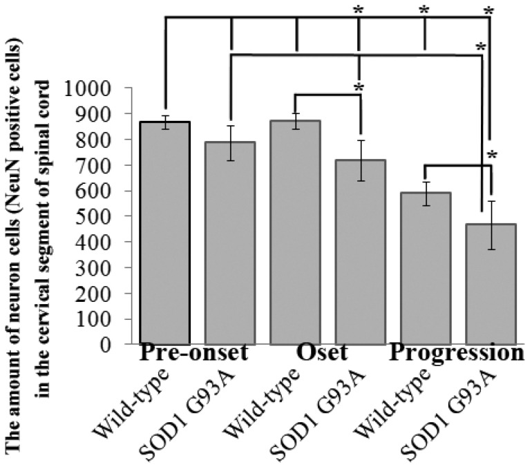
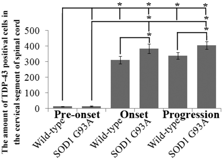
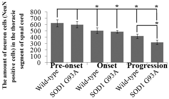
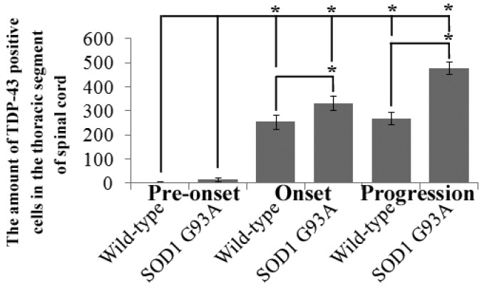
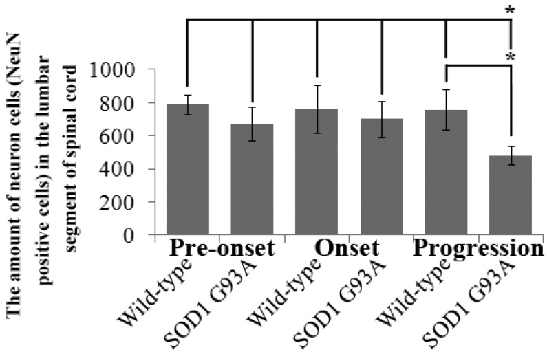
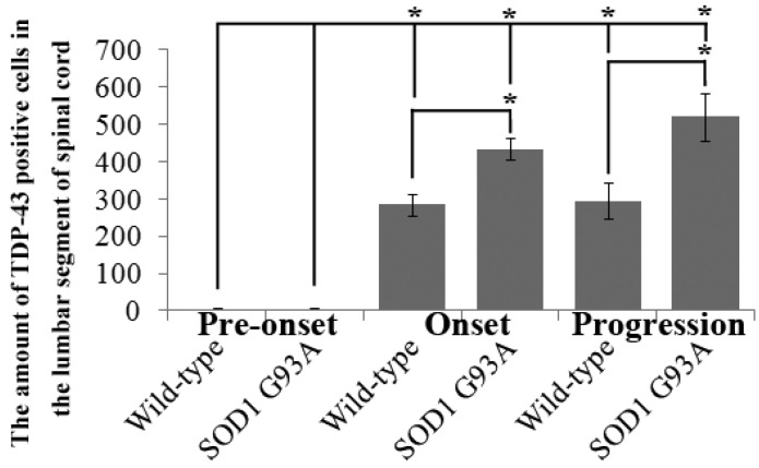
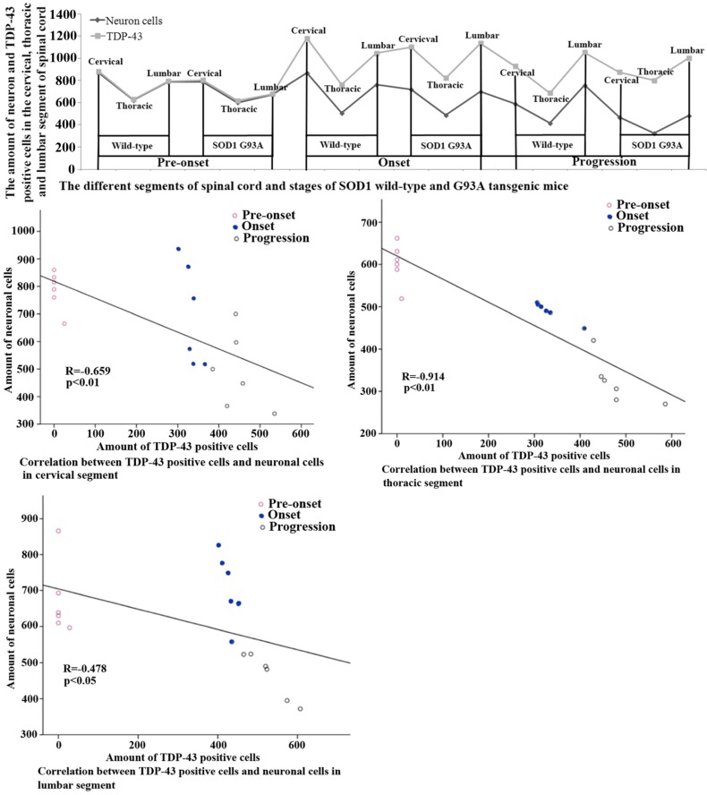
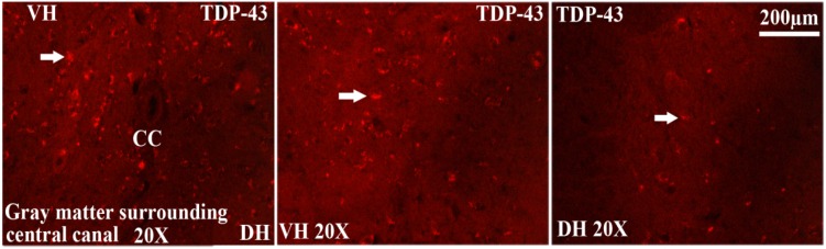
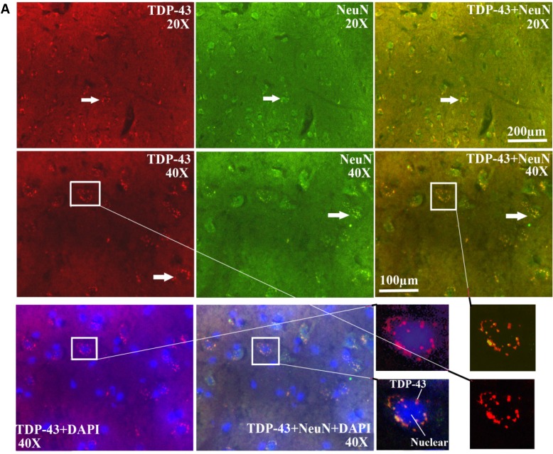
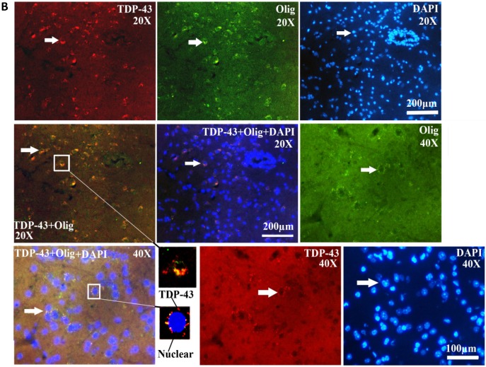
The recent investigation suggested that the TDP-43 protein was closely related to the motor neuron degeneration in amyotrophic lateral sclerosis (ALS), but the pathogenesis contributed to motor neuron degeneration largely remained unknown. Therefore, we detected the alteration of TDP-43 expression and distribution in the adult spinal cord of the SOD1 G93A transgenic mouse model for searching the possible pathogenesis of ALS. We examined the TDP-43 expression and distribution in the different anatomic regions, segments and neural cells in the adult spinal cord at the different stages of the SOD1 wild-type and G93A transgenic model by the fluorescent immunohistochemical technology. We revealed that the amount of TDP-43 positive cell was cervical>lumbar>thoracic segment, that in the ventral horn was more than that in the dorsal horn, a few of TDP-43 protein sparsely expressed and distributed in the other regions, the TDP-43 protein weren't detected in the white matter and the central canal. The TDP-43 protein was mostly expressed and distributed in the nuclear of neuron cells and the cytoplasm of oligodendrocyte cells of the gray matter surrounding the central canal of spinal cord by the granular shape in the SOD1 wild-type and G93A transgenic mice. The amount of TDP-43 positive cell significantly increased at the onset and progression stages of ALS following with the increase of neuron death in spinal cord, particularly in the ventral horn of cervical segment at the progression stage. Our results suggested that the overexpression of TDP-43 protein in the neuron and oligodendrocyte cell causes the progressive motor neuron degeneration in the ALS-like mouse model.

Keywords: Amyotrophic lateral sclerosis. Animal models. Mechanism of neurodegenerative diseases. Motor neuron diseases. Motor neuron. Neurodegeneration. Neurodegenerative disease. SOD1. Transgenic mice..


Conflict of interest statement

The authors have declared that no competing interest exists.

Figures

Figure 1
Figure 1
The amount of neuron cells (NeuN positive cells) in the cervical segment of spinal cord at the different stages of the SOD1 wild-type and G93A transgenic mice. In the SOD1 wild-type mice, the neuron cells weren't significantly different between the period of the pre-onset (60-70 days) and the onset (90-100 days) stages of ALS, but the neuron cells significantly decreased at the period of the progression stage (120-130 days) (*p<0.05). In the SOD1 G93A mice, the neuron cells gradually decreased from the pre-onset to the progression stages of ALS, but the neuron cells significantly decreased compared with the SOD1 wild-type mice at the onset and progression stages (*p<0.05). The result indicated that the neuron cells existed a gradual decrease tendency from 60 to 130 days, and the neuron cells significantly decreased after 120 days in the normal life-span of mice, but the neuron cells significantly decreased from 90 days in the SOD1 G93A mice. The decrease of neuron cells in the SOD1 G93A mice was significantly earlier than that in the wild-type mice (*p<0.05). The significant death of neuron cells began from the onset stage of ALS in the cervical segment of spinal cord.
Figure 2
Figure 2
The amount of TDP-43 positive cells in the cervical segment of spinal cord at the different stages of the SOD1 wild-type and G93A transgenic mice. The TDP-43 amount significantly increased from 90 days of the SOD1 wild-type and G93A transgenic mice (*p<0.05), and the increase of TDP-43 amount in the SOD1 G93A mice was more significant than that in wild-type mice (*p<0.05). The result indicated that the abnormal increase of TDP-43 protein mainly occurred at the onset and progression stages during the development of ALS in the cervical segment of spinal cord.
Figure 3
Figure 3
The amount of neuron cells in the thoracic segment of spinal cord at the different stages of the SOD1 wild-type and G93A transgenic mice. The neuron cells significantly decreased from the period of onset stages of ALS in the SOD1 wild-type and G93A transgenic mice (*p<0.05), and the decrease of neuron cells at the progression of SOD1 G93A mice was more significant than that at the pre-set and onset stages (*p<0.05). The result indicated that the significant death of neuron cells mainly occurred at the progression stage of ALS in the thoracic segment of spinal cord.
Figure 4
Figure 4
The amount of TDP-43 positive cells in the thoracic segment of spinal cord at the different stages of the SOD1 wild-type and G93A transgenic mice. There almost weren't the TDP-43 positive cells distribution in the period of pre-onset of the SOD1 wild-type and G93A transgenic mice. The TDP-43 positive cells dramatically increased at the period of the onset and progression stages in the SOD1 wild-type and G93A transgenic mice, the increase of TDP-43 in the SOD1 G93A transgenic mice was more significant than that in the wild-type mice. The TDP-43 protein gradually increased at the period of the onset and progression stages of both the SOD1 wild-type and G93A transgenic mice, but the increase of TDP-43 protein in the G93A transgenic mice was more significant than that in the wild-type mice. The results indicated that the abnormal increase of TDP-43 took part at the onset and progression during the development of ALS in the thoracic segment of spinal cord.
Figure 5
Figure 5
The amount of neuron cells in the lumbar segment of spinal cord at the different stages of the SOD1 wild-type and G93A transgenic mice. The neuron cells significantly decreased at the progression of the SOD1 G93A transgenic mice, the amount of neuron cells weren't any significant difference at the period of the pre-onset and onset stages of the SOD1 wild-type and G93A transgenic mice. The results indicated that the significant death of neuron cells mainly occurred at the progression stages of ALS in the lumbar segment of spinal cord.
Figure 6
Figure 6
The amount of TDP-43 positive cells in the lumbar segment of spinal cord at the different stages of the SOD1 wild-type and G93A transgenic mice. There almost weren't the TDP-43 positive cells distribution in the pre-onset of the SOD1 wild-type and G93A transgenic mice. The TDP-43 positive cells dramatically increased at the period of the onset and progression stages in the SOD1 wild-type and G93A transgenic mice, the increase of TDP-43 in the SOD1 G93A transgenic mice was more significant than that in the wild-type mice. The results indicated that the abnormal increase of TDP-43 participated in the onset and progression stages during the development of ALS in the lumbar segment of spinal cord.
Figure 7
Figure 7
The relationship between the neuron cells decrease and the TDP-43 protein increase in the cervical, thoracic and lumbar segments of spinal cord at the different stages of the SOD1 wild-type and G93A transgenic mice. The increase of neuron death was consistent with the increase of TDP-43 protein in the cervical, thoracic and lumbar segment of spinal cord and at the onset and progression stages of the SOD1 wild-type and G93A transgenic mice, and the relationship between the neuron cells decrease and the TDP-43 protein increase in the SOD1 G93A transgenic mice was particularly significant compared with that of the wild-type transgenic mice. The results indicated that the increase of TDP-43 protein contributed to the progressive neuron death in the development of ALS.
Figure 8
Figure 8
The TDP-43 expression and distribution in the different regions of spinal cord at the different stages of the SOD1 wild-type and G93A transgenic mice. The TDP-43 positive cells mainly were distributed in the gray matter surrounding the central canal of spinal cord, that in the ventral horn was more than that in the dorsal horn of spinal cord. The expression and distribution of TDP-43 positive cells was closer to the central canal and the more intensive. The TDP-43 positive cells weren't detected in the central canal and the white matter of spinal cord. The Figure 8 was the representative images of the TDP-43 expression and distribution in the different regions of spinal cord at the different stages of the SOD1 wild-type and G93A transgenic mice. Abbreviation: VH=Ventral Horn; CC=Central Canal; DH=Dorsal Horn. White arrow indicates the positive cell.
Figure 9
Figure 9
The TDP-43 expression and distribution in the different neural cells at the different stages of the SOD1 wild-type and G93A transgenic mice. Almost all TDP-43 positive cells were distributed by the granular shape in the unclear of the neuron cells and the cytoplasm of oligodendrocyte cells in the SOD1 wild-type and G93A transgenic mice, the majority of TDP-43 positive cells were distributed in the neuron cells, the TDP-43 positive cells almost weren't detected in the astrocyte and microglia cell. The results indicated that the TDP-43 protein mainly expressed in the neuron cells and oligodendrocytes cells, particularly in the neuron cells. The Figure A and B were the representative images of the TDP-43 expression and distribution in the different neural cells at the different stages of the SOD1 wild-type and G93A transgenic mice.
Figure 9
Figure 9
The TDP-43 expression and distribution in the different neural cells at the different stages of the SOD1 wild-type and G93A transgenic mice. Almost all TDP-43 positive cells were distributed by the granular shape in the unclear of the neuron cells and the cytoplasm of oligodendrocyte cells in the SOD1 wild-type and G93A transgenic mice, the majority of TDP-43 positive cells were distributed in the neuron cells, the TDP-43 positive cells almost weren't detected in the astrocyte and microglia cell. The results indicated that the TDP-43 protein mainly expressed in the neuron cells and oligodendrocytes cells, particularly in the neuron cells. The Figure A and B were the representative images of the TDP-43 expression and distribution in the different neural cells at the different stages of the SOD1 wild-type and G93A transgenic mice.

Similar articles

Cited by

References

    1. Ou SH, Wu F, Harrich D, García-Martínez LF, Gaynor RB. Cloning and characterization of a novel cellular protein, TDP-43, that binds to human immunodeficiency virus type 1 TAR DNA sequence motifs. Journal of virology. 1995;69:3584–96. - PMC - PubMed
    1. Sephton CF, Cenik C, Kucukural A, Dammer EB, Cenik B, Han Y. et al. Identification of neuronal RNA targets of TDP-43-containing ribonucleoprotein complexes. Journal of biological chemistry. 2011;286:1204–1215. - PMC - PubMed
    1. Kuo PH, Doudeva LG, Wang YT, Shen CK, Yuan HS. Structural insights into TDP-43 in nucleic-acid binding and domain interactions. Nucleic acids research. 2009;37:1799–808. - PMC - PubMed
    1. Strong MJ, Volkening K, Hammond R, Yang W, Strong W, Leystra-Lantz C. et al. TDP43 is a human low molecular weight neurofilament (hNFL) mRNA-binding protein. Molecular and cellular neuroscience. 2007;35:320–7. - PubMed
    1. Wang IF, Wu LS, Chang HY, Shen CK. TDP-43, the signature protein of FTLD-U, is a neuronal activity-responsive factor. Journal of neurochemistry. 2008;105:797–806. - PubMed

Publication types

MeSH terms