. 2016 Jul 18;6(11):1833-43.
doi: 10.7150/thno.16047. eCollection 2016.

Polydopamine Coated Single-Walled Carbon Nanotubes as a Versatile Platform with Radionuclide Labeling for Multimodal Tumor Imaging and Therapy

Affiliations

Polydopamine Coated Single-Walled Carbon Nanotubes as a Versatile Platform with Radionuclide Labeling for Multimodal Tumor Imaging and Therapy

He Zhao et al. Theranostics. .

Abstract

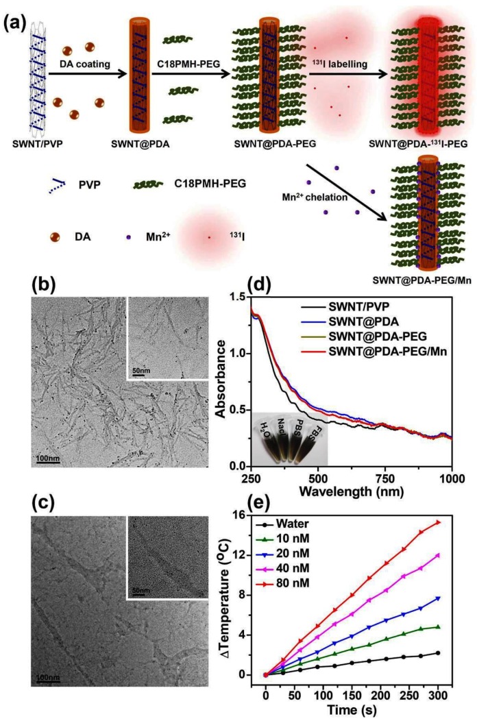
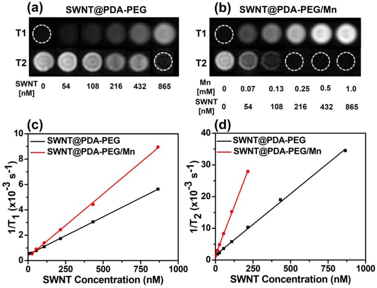
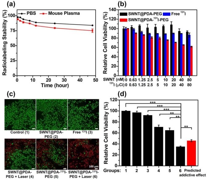
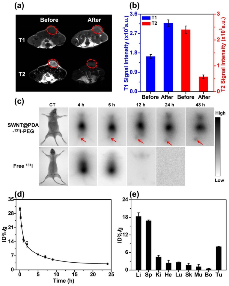
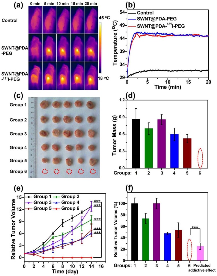
Single-walled carbon nanotubes (SWNTs) with various unique properties have attracted great attention in cancer theranostics. Herein, SWNTs are coated with a shell of polydopamine (PDA), which is further modified by polyethylene glycol (PEG). The PDA shell in the obtained SWNT@PDA-PEG could chelate Mn(2+), which together with metallic nanoparticulate impurities anchored on SWNTs offer enhanced both T1 and T2 contrasts under magnetic resonance (MR) imaging. Meanwhile, also utilizing the PDA shell, radionuclide (131)I could be easily labeled onto SWNT@PDA-PEG, enabling nuclear imaging and radioisotope cancer therapy. As revealed by MR & gamma imaging, efficient tumor accumulation of SWNT@PDA-(131)I-PEG is observed after systemic administration into mice. By further utilizing the strong near-infarared (NIR) absorbance of SWNTs, NIR-triggered photothermal therapy in combination with (131)I-based radioisotope therapy is realized in our animal experiments, in which a remarkable synergistic antitumor therapeutic effect is observed compared to monotherapies. Our work not only presents a new type of theranostic nanoplatform based on SWNTs, but also suggests the promise of PDA coating as a general approach to modify nano-agents and endow them with highly integrated functionalities.

Keywords: Combined therapy; Multimodal imaging; Polydopamine; Radiolabeling; Single-walled carbon nanotubes.


Conflict of interest statement

The authors have declared that no competing interest exists.

Figures

Figure 1
Figure 1
Preparation and characterization of SWNT@PDA-PEG. (a) A schematic illustration for the fabrication of SWNT@PDA-PEG. (b&c) TEM images of SWNT/PVP (b) and SWNT@PDA (c). Inset is a TEM image with higher resolution. (d) UV-Vis-NIR absorbance spectra of SWNT/PVP, SWNT@PDA, SWNT@PDA-PEG and SWNT@PDA-PEG/Mn solutions at the same SWNT concentration. Inset: a photo of SWNT@PDA-PEG/Mn in various types of physiological buffers. (e) Temperature change curves of SWNT@PDA-PEG solution at different concentrations (i.e. 10, 20, 40, 80 nM) exposed to the 808 nm laser with a power density of 0.7 W cm-2 for 5 min. The molar concentrations of SWNTs were determined by a literature method.
Figure 2
Figure 2
MR imaging of SWNT@PDA-PEG samples without or with chelation of Mn2+. (a&b) T1- and T2- weighted MR images of SWNT@PDA-PEG samples before (a) and after (b) Mn2+ chelating. The SWNT concentrations varied from 0 to 865 nM, corresponding to Mn concentrations from 0 to 1 mM (for SWNT@PDA-PEG/Mn). (c&d) T1 (c) and T2 (d) relaxation rates of SWNT@PDA-PEG and SWNT@PDA-PEG/Mn at different SWNT concentrations.
Figure 3
Figure 3
In vitro cell experiments. (a) The radiolabeling stability of SWNT@PDA-131I-PEG incubated with PBS and mouse plasma at 37 °C. (b) The relative viabilities of 4T1 cells incubated with various concentrations of SWNT@PDA-PEG, free 131I and SWNT@PDA-131I-PEG. For SWNT@PDA-PEG and SWNT@PDA-131I-PEG, SWNT concentrations varied from 0 to 80 nM. For free 131I and SWNT@PDA-131I-PEG, 131I concentrations varied from 0 to 80 μCi. (c&d) The fluorescence imaging of Calcein AM/PI stained 4T1 cells (c) and their relative viabilities (d) after different treatments. Group 1: Control; Group 2: SWNT@PDA-PEG; Group 3: Free 131I; Group 4: SWNT@PDA-PEG + Laser; Group 5: SWNT@PDA-131I-PEG; Group 6: SWNT@PDA-131I-PEG + Laser. The predicted addictive effect was calculated by multiplying the remained cell viability ratio in group 4 (PTT only) with that in group 5 (RIT only). The concentration of 131I was 150 μCi (corresponding to 80 nM of SWNT@PDA-PEG in the SWNT@PDA-131I-PEG sample). The laser irradiation was conducted at about 0.3 W cm-2 to maintain the temperature at about 46 oC for 20 min. P values were calculated by the Student's two-tailed t-test (*** p <0.001 or ** p <0.01).
Figure 4
Figure 4
In vivo behaviors of SWNT@PDA-131I-PEG. (a) In vivo T1- (upper) and T2-weighted MR images (bottom) of a 4T1 tumor-bearing mouse taken before injection (left) and 24 h post injection (right) of SWNT@PDA-PEG/Mn. Obvious brightening and darkening effects showed up in the tumor after i.v. injection of SWNT@PDA-PEG/Mn by T1- and T2-weighted MR imaging, respectively. (b) T1- (left) and T2-weighted (right) MR signals intensities of the tumor before and 24 h post i.v. injection of SWNT@PDA-PEG/Mn, which offered strong tumor contrasts under both T1- and T2-weighted MR imaging modes. (c) Gamma imaging of SWNT@PDA-131I-PEG-treated mice and free 131I-treated mice. Notably, free 131I was completely excreted after 12 h, while SWNT@PDA-131I-PEG showed obvious tumor accumulation. (d) The blood circulation of SWNT@PDA-131I-PEG after i.v. injection determined by gamma-counting. (e) The biodistribution of SWNT@PDA-131I-PEG in 4T1 tumor-bearing mice measured at 24 h p.i. Li: liver; Sp: spleen; Ki: kidney; He: heart; Lu: lung; Sk: skin; Mu: muscle; Bo: bone; Tu: tumor.
Figure 5
Figure 5
In vivo combined therapy with SWNT@PDA-131I-PEG. (a) IR thermal images of 4T1 tumor-bearing mice with or without injection of SWNT@PDA-131I-PEG ([SWNT]:4.3 mg/kg, 200 μCi of 131I per mouse), under the 808 nm laser irradiation (about 0.6 W cm-2). (b) Temperature change curves of tumors monitored by the IR thermal camera during laser irradiation. (c) Photos of the tumors collected from different groups of mice at day 14 after different treatments. Note that in the combined therapy group, all of the tumors were completely eliminated after treatment. (d) The tumors collected from all the groups were weighted 14 days after various treatments. (e) Tumor volume growth curves of different groups of mice after various treatments. The temperature was kept at about 46 oC for 20 minutes by NIR irradiation. (f) Relative tumor volume of different groups after various treatments. Group 1: Control; Group 2: SWNT@PDA-PEG; Group 3: Free 131l; Group 4: SWNT@PDA-PEG + Laser; Group 5: SWNT@PDA-131I-PEG; Group 6: SWNT@PDA-131I-PEG + Laser. The predicted addictive effect was calculated by multiplying the relative tumor volume ratio in group 4 (PTT only) with that in group 5 (RIT only). Statistical analysis was performed using the Student's two-tailed t-test (*** p <0.001).
Figure 6
Figure 6
H&E staining and TUNEL staining of tumor slices. (a) TUNEL staining of the tumor slices taken at day 2 post the initiation of treatments. The combined internal RT with PTT offered remarkable synergistic therapeutic effect in inhibiting the tumor growth. (b) H&E stained images of the tumor slices taken at day 2 post the initiation of treatments. Group 1: Control; Group 2: SWNT@PDA-PEG; Group 3: Free 131l; Group 4: SWNT@PDA-PEG + Laser; Group 5: SWNT@PDA-131I-PEG; Group 6: SWNT@PDA-131I-PEG + Laser.

References

    1. Chen Z-Y, Wang Y-X, Lin Y, Zhang J-S, Yang F, Zhou Q-L, Advance of molecular imaging technology and targeted imaging agent in imaging and therapy. BioMed research international. 2014; 2014. - PMC - PubMed
    1. Liu T, Shi S, Liang C, Shen S, Cheng L, Wang C. et al. Iron oxide decorated MoS2 nanosheets with double PEGylation for chelator-free radiolabeling and multimodal imaging guided photothermal therapy. ACS nano. 2015;9:950–60. - PMC - PubMed
    1. Zhong J, Wen L, Yang S, Xiang L, Chen Q, Xing D. Imaging-guided high-efficient photoacoustic tumor therapy with targeting gold nanorods. Nanomedicine: Nanotechnology, Biology and Medicine. 2015;11:1499–509. - PubMed
    1. Zhong J, Yang S, Wen L, Xing D. Imaging-guided photoacoustic drug release and synergistic chemo-photoacoustic therapy with paclitaxel-containing nanoparticles. Journal of Controlled Release. 2016;226:77–87. - PubMed
    1. Xu X, Ho W, Zhang X, Bertrand N, Farokhzad O. Cancer nanomedicine: from targeted delivery to combination therapy. Trends in molecular medicine. 2015;21:223–32. - PMC - PubMed