. 2016 Jun;9(2):258-267.
doi: 10.1007/s12195-016-0437-8. Epub 2016 Apr 18.

Nuclear lamins in cancer

Affiliations

Nuclear lamins in cancer

Jerome Irianto et al. Cell Mol Bioeng. 2016 Jun.

Abstract

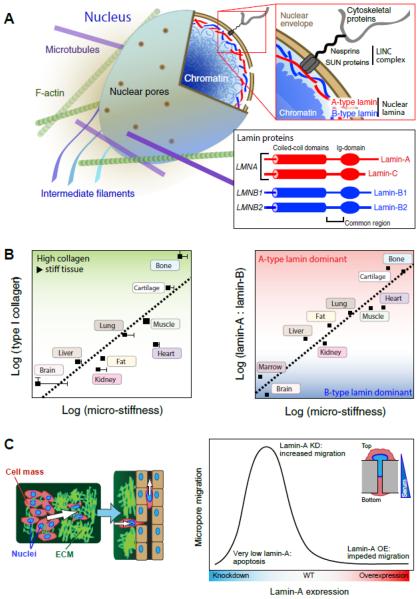
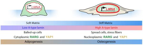
Dysmorphic nuclei are commonly seen in cancers and provide strong motivation for studying the main structural proteins of nuclei, the lamins, in cancer. Past studies have also demonstrated the significance of microenvironment mechanics to cancer progression, which is extremely interesting because the lamina was recently shown to be mechanosensitive. Here, we review current knowledge relating cancer progression to lamina biophysics. Lamin levels can constrain cancer cell migration in 3D and thereby impede tumor growth, and lamins can also protect a cancer cell's genome. In addition, lamins can influence transcriptional regulators (RAR, SRF, YAP/TAZ) and chromosome conformation in lamina associated domains. Further investigation of the roles for lamins in cancer and even DNA damage may lead to new therapies or at least to a clearer understanding of lamins as bio-markers in cancer progression.

Keywords: LADs; Nuclear lamina; SRF; YAP/TAZ; cancer; homeostasis; mechanotransduction.


Figures

Fig. 1
Fig. 1. Nuclear lamin levels scale with matrix stiffness and dictate cancer cell migration potential
(A) A-type and B-type lamins form juxtaposed networks on the inside of the nuclear envelope; they are effectively located at an interface between chromatin and the cytoskeleton, to which the lamina is attached through the `LINC' (linker of nucleo- and cytoskeleton) complex. `A-type lamins', lamins A and C are alternative spliceoform products of the LMNA gene; `B-type lamins', lamins B1 and B2 are protein products of LMNB1 and LMNB2 respectively (adapted from ©Buxboim et al. 2010, originally published in The Journal of Cell Science). (B - left) The quantity of collagen-1 present in tissues scales with tissue micro-elasticity. As collagen is one of the most prevalent proteins in the body, it is perhaps expected that it defines mechanical properties. (B - right) The composition of the nuclear lamina scales with tissue microelasticity. A-type lamins dominate the lamina in stiff tissue, whereas B-type lamins are prevalent in soft tissue. (C - left) As the largest and stiffest organelle in the cell, the nucleus can act as an `anchor', preventing cell movement through the matrix or into surrounding vasculature. (C - right) As a model of migration through matrix, cells are induced to pass through 3 μm-pores, a diameter sufficiently small to require deformation of the nucleus (inset). Lamin-A overexpression inhibits migration, whereas knockdown increases migration, up to a point at which significant apoptosis is observed. Thus extremely low or high lamin-A,C levels are unfavorable for cell migration, an observation with potential impact on understanding of processes such as cell migration during development and cancer metastasis.
Fig. 2
Fig. 2. Decisions of cell fate downstream of lamin-A,C regulation
MSCs cultured on soft and stiff substrates take on differing phenotypes and favor alternate cell fates. On soft substrate, MSCs exhibit small nuclear and cellular spread areas, and the nuclear lamina is thinned by a stress-sensitive phosphorylation feedback mechanism. The transcription factors RARG and YAP1 remain in the cytoplasm, and adipogenic cell fate is preferred. Conversely, on stiff substrate, cells spread extensively with nuclei that are pinned down by well-developed stress fibers. Lamin-A,C is less phosphorylated under strain, thus strengthening the lamina; RARG also translocates to the nucleus, increasing LMNA transcription. Activity of the transcription factor SRF (downstream of lamin-A,C) increases expression of cytoskeletal components. Under these conditions, YAP1 translocates to the nucleus and cells favor osteogenesis. On both soft and stiff substrates, the effects of matrix elasticity and lamin level cooperate to enhance differentiation: lamin-A,C knockdown on soft matrix leads to more adipogenesis; lamin-A overexpression on stiff matrix leads to more osteogenesis.

References

    1. Agrelo R, Setien F, Espada J, Artiga MJ, Rodriguez M, Perez-Rosado A, Sanchez-Aguilera A, Fraga MF, Piris MA, Esteller M. Inactivation of the lamin A/C gene by CpG island promoter hypermethylation in hematologic malignancies, and its association with poor survival in nodal diffuse large B-cell lymphoma. Journal of clinical oncology: official journal of the American Society of Clinical Oncology. 2005;23(17):3940–7. - PubMed
    1. Akhtar W, de Jong J, Pindyurin AV, Pagie L, Meuleman W, de Ridder J, Berns A, Wessels LFA, van Lohuizen M, van Steensel B. Chromatin Position Effects Assayed by Thousands of Reporters Integrated in Parallel. Cell. 2013;154(4):914–927. - PubMed
    1. Alfonso P, Canamero M, Fernandez-Carbonie F, Nunez A, Casal JI. Proteome analysis of membrane fractions in colorectal carcinomas by using 2D-DIGE saturation labeling. Journal of proteome research. 2008;7(10):4247–55. - PubMed
    1. Amendola M, van Steensel B. Nuclear lamins are not required for lamina-associated domain organization in mouse embryonic stem cells. EMBO reports. 2015;16(5):610–7. - PMC - PubMed
    1. Aragona M, Panciera T, Manfrin A, Giulitti S, Michielin F, Elvassore N, Dupont S, Piccolo S. A mechanical checkpoint controls multicellular growth through YAP/TAZ regulation by actin-processing factors. Cell. 2013;154(5):1047–59. - PubMed

LinkOut - more resources