. 2016;6(43):36794-36802.
doi: 10.1039/C6RA05895G. Epub 2016 Mar 30.

Single Nanoparticle Plasmonic Spectroscopy for Study of the Efflux Function of Multidrug ABC Membrane Transporters of Single Live Cells

Affiliations

Single Nanoparticle Plasmonic Spectroscopy for Study of the Efflux Function of Multidrug ABC Membrane Transporters of Single Live Cells

Lauren M Browning et al. RSC Adv. 2016.

Abstract

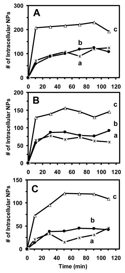
ATP-binding cassette (ABC) membrane transporters exist in all living organisms and play key roles in a wide range of cellular and physiological functions. The ABC transporters can selectively extrude a wide variety of structurally and functionally unrelated substrates, leading to multidrug resistance. Despite extensive study, their efflux molecular mechanisms remain elusive. In this study, we synthesized and characterized purified silver nanoparticles (Ag NPs) (97 ± 13 nm in diameter), and used them as photostable optical imaging probes to study efflux kinetics of ABC membrane transporters (BmrA) of single live cells (B. subtillis). The NPs with concentrations up to 3.7 pM were stable (non-aggregated) in a PBS buffer and biocompatible with the cells. We found a high dependence of accumulation of the intracellular NPs in single live cells (WT, Ct-BmrA-EGFP, ΔbmrA) upon the cellular expression level of BmrA and NP concentration (0.93, 1.85 and 3.7 pM), showing the highest accumulation of intracellular NPs in ΔbmrA (deletion of BmrA) and the lowest ones in Ct-BmrA-EGFP (over-expression of BmrA). Interestingly, the accumulation of intracellular NPs in ΔbmrA increases nearly proportionally with the NP concentration, while those in WT and Ct-BrmA-EGFP do not. This suggests that the NPs enter the cells via passive diffusion driven by concentration gradients and are extruded out of cells by BmrA transporters, similar to conventional pump substrates (antibiotics). This study shows that such large substrates (84-100 nm NPs) can enter into the live cells and be extruded out of the cells by BmrA, and the NPs can serve as nm-sized optical imaging probes to study the size-dependent efflux kinetics of membrane transporters in single live cells in real time.

Keywords: ABC (BmrA) transporter; Ag nanoparticle; Bacillus subtilis; multidrug resistance; single cell imaging; single nanoparticle plasmonic spectroscopy.


Figures

Figure 1
Figure 1
Characterization of size and optical properties of single Ag NPs. (A) HRTEM images of single Ag NPs shows ellipse shaped NPs. (B) Histograms of the size distribution of single Ag NPs measured by HRTEM shows an average diameter of 97 ± 13 nm, which was determined by averaging the length and width of each NP. (C) Representative dark-field optical image of single Ag NPs shows individual plasmonic green, green-yellow, yellow, and red NPs with (D) LSPR spectra at λmax (FWHM): (a) 521 (145), (b) 536 (118), (c) 560 (106), and (d) 657 (114) nm, respectively. Scale bars are 100 nm in (A) and 2 μm in (C). The scale bar in (C) shows the distances among single NPs, but not their sizes, due to optical diffraction limit.
Figure 2
Figure 2
Characterization of stability of Ag NPs (97 ± 13 nm) in the PBS buffer (0.5 mM phosphate buffer, 1.5 mM NaCl, pH = 7.0). (A) UV–visible absorption spectra (CNPs = 3.7 pM) of the Ag NPs at (a) 0 h and (b) 24 h, show peak absorbance of 1.9 at 505 nm (FWHM = 150 nm) and a shoulder peak with a peak absorbance of 1.6 at 419 nm (FWHM = 93 nm). (B) Sizes of NPs in the PBS buffer measured using DLS show average diameters of (a) 89 ± 17 nm at 0 h and (b) 92 ± 19 nm at 24 h, indicating that the sizes of Ag NPs in the PBS buffer remain essentially unchanged and they are stable in PBS buffer for 24 h.
Figure 3
Figure 3
Imaging of single intracellular and extracellular Ag NPs in single live cells using DFOMS. (A) Representative images of individual WT cells incubated with 3.7 pM Ag NPs show: (a) intracellular and extracellular NPs. (B) Zoom-in images of single cells show (a) intracellular and (b) extracellular NPs. (C) LSPR spectra of single NPs show peak wavelength (FWHM) at (a) 540 (96), (b) 552 (92) and (c) 578 (137) nm. The plasmonic colors of the NPs in (B) were characterized using their LSPR spectra in (C) and their pseudo colors were added into the images based upon their spectra. The scale bar is 6 μm in (A) and 2 μm in (B). Representative images of single cells (Ct-BmrA-EGFP and ΔBmrA) incubated with the NPs are shown in the Supporting Information (SI).
Figure 4
Figure 4
Study of dependence of accumulation and efflux kinetics of single Ag NPs in single live cells on the expression level of BmrA. Plots of number of intracellular NPs in (a) WT (ӿ), (b) Ct-BmrA-EGFP (•) and (c) ΔBmrA (Δ) cells that were incubated with (A) 3.7, (B) 1.85, and (C) 0.93 pM NPs over 2 h. The points represent the sum of three repeated experiments and the lines are added to guide the trend. At each point (every 20 min), 300 cells are analyzed.
Figure 5
Figure 5
Study of concentration-dependent accumulation and efflux kinetics of single Ag NPs in single live cells. Plots of number of intracellular NPs in (A) WT, (B) Ct-BmrA-EGFP and (C) ΔBmrA cells that were incubated with (a) 3.7, (b) 1.85, and (c) 0.93 pM NPs for 2 h. The points represent the sum of three repeated experiments and the lines are added to guide the trend. At each point (every 20 min), 300 cells are analyzed.
Figure 6
Figure 6
Characterization of the viability of single cells using live/dead bacLight viability and counting assay. (A) Dark field optical image and (B) fluorescence image of single (a) WT, (b) Ct-BmrA-EGFP, and (c) ΔBmrA cells that had been incubated with 3.7 pM NPs for 2 h show that the cells with or without intracellular NPs emit the green fluorescence of SYTO9 (λmax = 520 nm), but not red fluorescence, indicating that they are viable. (C) Plots of percentage of viable cells incubated with NPs for 2 h show that 97–98% of the cells (WT, Ct-BmrA-EGF or ΔBmrA) are alive. The scale bar is 8 μm.

Similar articles

Cited by

References

    1. Cole SP. J. Biol. Chem. 2014;289:30880–30888. - PMC - PubMed
    1. Nigam SK. Nat. Rev. Drug Discov. 2015;14:29–44. - PMC - PubMed
    1. Gottesman MM, Ambudkar SV, Xia D. Nat. Biotechnol. 2009;27:546–547. - PMC - PubMed
    1. Higgins CF. Res. Microbiol. 2001;152:205–210. - PubMed
    1. Hung LW, Wang IX, Nikaido K, Liu PQ, Ames GF, Kim SH. Nature. 1998;396:703–707. - PubMed

LinkOut - more resources