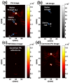
In vivo demonstration of reflection artifact reduction in photoacoustic imaging using synthetic aperture photoacoustic-guided focused ultrasound (PAFUSion)
- PMID: 27570690
- PMCID: PMC4986806
- DOI: 10.1364/BOE.7.002955
In vivo demonstration of reflection artifact reduction in photoacoustic imaging using synthetic aperture photoacoustic-guided focused ultrasound (PAFUSion)
Abstract
Reflection artifacts caused by acoustic inhomogeneities are a critical problem in epi-mode biomedical photoacoustic imaging. High light fluence beneath the probe results in photoacoustic transients, which propagate into the tissue and reflect back from echogenic structures. These reflection artifacts cause problems in image interpretation and significantly impact the contrast and imaging depth. We recently proposed a method called PAFUSion (Photoacoustic-guided focused ultrasound) to identify such reflection artifacts in photoacoustic imaging. In its initial version, PAFUSion mimics the inward-travelling wavefield from small blood vessel-like PA sources by applying ultrasound pulses focused towards these sources, and thus provides a way to identify the resulting reflection artifacts. In this work, we demonstrate reduction of reflection artifacts in phantoms and in vivo measurements on human volunteers. In view of the spatially distributed PA sources that are found in clinical applications, we implemented an improved version of PAFUSion where photoacoustic signals are backpropagated to imitate the inward travelling wavefield and thus the reflection artifacts. The backpropagation is performed in a synthetic way based on the pulse-echo acquisitions after transmission on each single element of the transducer array. The results provide a direct confirmation that reflection artifacts are prominent in clinical epi-photoacoustic imaging, and that PAFUSion can strongly reduce these artifacts to improve deep-tissue photoacoustic imaging.
Keywords: (170.3880) Medical and biological imaging; (170.5120) Photoacoustic imaging; (170.7170) Ultrasound.
Figures


References
-
- Xu M., Wang L. V., “Photoacoustic imaging in biomedicine,” Rev. Sci. Instrum. 77, 041101 (2006).
-
- Dean-Ben X. L., Razansky D., “Adding fifth dimension to optoacoustic imaging: volumetric time-resolved spectrally enriched tomography,” Light Sci. Appl. 3, e137 (2014).
LinkOut - more resources
Full Text Sources
Other Literature Sources
