. 2016 Jul 27;3(4):ENEURO.0029-16.2016.
doi: 10.1523/ENEURO.0029-16.2016. eCollection 2016 Jul-Aug.

Axonal Localization of Integrins in the CNS Is Neuronal Type and Age Dependent

Affiliations

Axonal Localization of Integrins in the CNS Is Neuronal Type and Age Dependent

Melissa R Andrews et al. eNeuro. .

Abstract

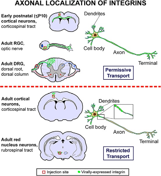
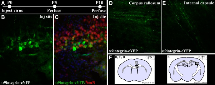
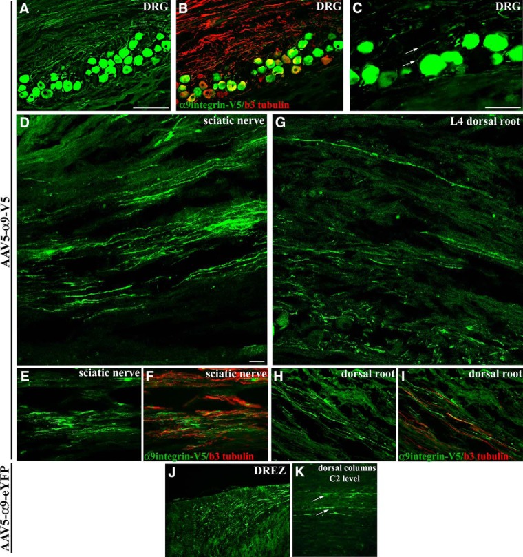
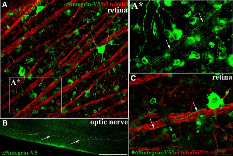
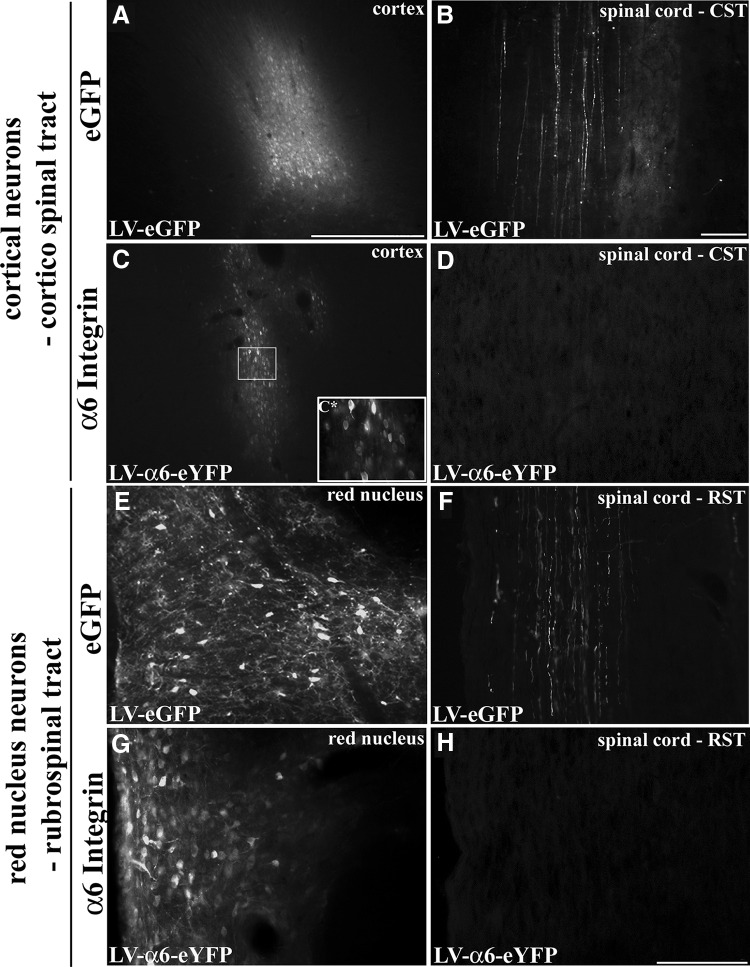
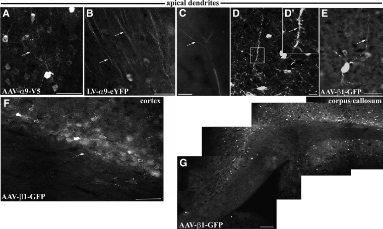
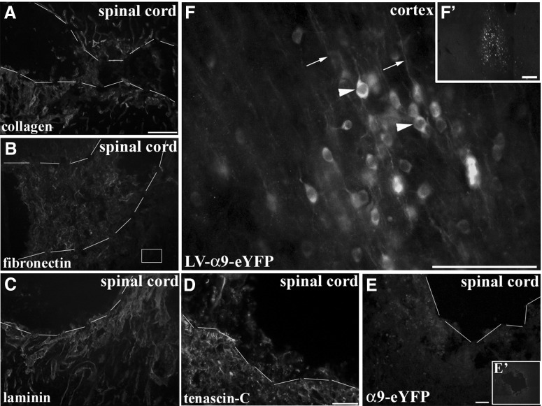
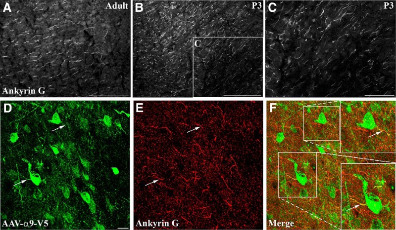
The regenerative ability of CNS axons decreases with age, however, this ability remains largely intact in PNS axons throughout adulthood. These differences are likely to correspond with age-related silencing of proteins necessary for axon growth and elongation. In previous studies, it has been shown that reintroduction of the α9 integrin subunit (tenascin-C receptor, α9) that is downregulated in adult CNS can improve neurite outgrowth and sensory axon regeneration after a dorsal rhizotomy or a dorsal column crush spinal cord lesion. In the current study, we demonstrate that virally expressed integrins (α9, α6, or β1 integrin) in the adult rat sensorimotor cortex and adult red nucleus are excluded from axons following neuronal transduction. Attempts to stimulate transport by inclusion of a cervical spinal injury and thus an upregulation of extracellular matrix molecules at the lesion site, or cotransduction with its binding partner, β1 integrin, did not induce integrin localization within axons. In contrast, virally expressed α9 integrin in developing rat cortex (postnatal day 5 or 10) demonstrated clear localization of integrins in cortical axons revealed by the presence of integrin in the axons of the corpus callosum and internal capsule, as well as in the neuronal cell body. Furthermore, examination of dorsal root ganglia neurons and retinal ganglion cells demonstrated integrin localization both within peripheral nerve as well as dorsal root axons and within optic nerve axons, respectively. Together, our results suggest a differential ability for in vivo axonal transport of transmembrane proteins dependent on neuronal age and subtype.

Keywords: adeno-associated virus; axon initial segment; dorsal root ganglia; integrin; retinal ganglion cell; sensorimotor cortex.


Figures

None
Graphical abstract
Figure 1.
Figure 1.
eYFP-tagged α9 integrin is transported into early postnatal cortical axons. Schematic of experimental design indicating the ages at which the lentivirus was injected and the time points of perfusion and tissue analysis (A). Fluorescent images of cortical injection site 5 d following LV-PGK- α9integrin-eYFP injection showing eYFP-labeled cortical neurons (green; B) colabeled with NeuN (red) and bisbenzimide nuclear label (blue; C). Fluorescent images of eYFP-labeled α9integrin within axons of the corpus callosum (D) or internal capsule (E), 5 or 10 d following cortical injection, respectively. F, Coronal brain illustrations (left) indicate approximate area of injection site (of boxed area for B, C) and area of image of corpus callosum (in D) and (right) indicate approximate area of image of internal capsule (in E). Scale bars: B, C, 100 μm; D, E, 50 μm.
Figure 2.
Figure 2.
V5 and eYFP-tagged α9 integrin expressed in DRG neurons is transported to the central and peripheral branches of DRG axons. DRG neurons express α9 integrin-V5 (green in all panels) 4 weeks following injection of AAV5-CAG-α9integrin-V5 into the L4 and L5 DRG (A) including within the proximal neuronal processes (C, arrows), shown colabeled with β3 tubulin (red) (B). Confocal (D) and epifluorescent (E) images show V5-labeled α9 integrin within axons in the sciatic nerve, 4 weeks following DRG injection, colabeled with anti-β3 tubulin (red; F). Confocal (G) and epifluorescent (H) images show V5-labeled α9 integrin within axons in the dorsal root, 4 weeks following DRG injection, colabeled with anti-β3 tubulin (red) (I). Epifluorescent image of eYFP-labeled α9 integrin (J, K, green) in the axons of the dorsal root entry zone leading into the dorsal column, 6 weeks following DRG injection of AAV-CMV-α9integrin-eYFP into the C5 and C6 DRG (J), and in axons in the dorsal columns (K, arrows) observed in sagittal section at level C2 (K). Scale bars: A, B, J, K, 200 μm; C, E, F, H, I, 100 μm; D, G, 20 μm.
Figure 3.
Figure 3.
α9integrin-V5 expressed in adult RGCs is transported into optic nerve axons. Confocal images of flat mount retina show RGCs immunolabeled with anti-V5 (green) and colabeled with anti-β3 tubulin (red) 3 weeks after intravitreal injection of AAV2-CAG-α9integrin-V5 (A, C). A*, A high magnification image (from A) of integrin-containing axons in a fascicle (arrows) travelling toward the optic nerve. Epifluorescent images in B of optic nerve indicate V5-labeled α9integrin within axon fibers of the optic nerve 3 weeks following AAV injection. Arrows in C indicate V5-labeled axons following along the course of β3 tubulin axons. Scale bars: A, C, 20 μm; B, 50 μm.
Figure 4.
Figure 4.
α6Integrin expressed in adult cortical or rubrospinal neurons is not transported down CST or RST axons. Adult motor cortex 3 weeks following injection of LV-PGK-eGFP (A) or LV-PGK-α6integrin-eYFP (C). In cervical spinal cord, axons are filled with eGFP in the CST following LV-PGK-eGFP cortical injection (B), but no integrins are observed after LV-PGK-α6integrin-eYFP injection (D). C*, High magnification view of LV-α6 integrin transduced cortical neurons. Adult red nucleus 3 weeks following injection of LV-PGK-eGFP (E) or LV-PGK-α6integrin-eYFP (G). Within the cervical spinal cord, only in the LV-PGK-eGFP injected groups are RST fibers found labeled with GFP (F) and not following LV-PGK-α6integrin-eYFP injection (H). Scale bars: A, C, E, G, 500 μm; B, D, 100 μm; F, H, 200 μm.
Figure 5.
Figure 5.
α9Integrin- and β1integrin-transduced adult cortical neurons express integrins in their dendrites but not in their axons. α9- and β1-transduced neurons (AE) show prominent apical and basal dendrites (white arrows), but integrins have not entered the axon beyond the very proximal processes (A, E, F). B, A region of cortex above the injection site demonstrating α9-transduced neurons with YFP-immunopositive integrin within apical dendrites. C, Detail of YFP-immunopositive α9integrin dendritic arbors branching (white arrows) near the surface of the cortex. D, A confocal image of YFP-immunopositive α9integrin within a dendrite with prominent dendritic spines (white arrows). F, G, The base of the cortex and the underlying white matter 4 weeks following AAV5-β1-GFP cortical injections demonstrating transduction of neurons throughout a wide area of cortex. Most of the white matter is devoid of tagged integrin, but a few fine processes of neurons very close to the white matter can be seen, demonstrating that labeled processes in white matter can be seen if present. G, A composite showing subcortical white matter from the midline (left of picture) to lateral cortex. Although there are many integrin-transduced neurons in the overlying cortex, no integrin-containing axons are observed in the white matter of the corpus callosum. Scale bars: A, C, E, F, 50 μm; B, G, 100 μm; D, 10 μm.
Figure 6.
Figure 6.
CNS injury induces upregulation of ECM expression but does not induce integrin localization in adult CST axons. Cervical dorsal column crush lesion leads to upregulation of ECM molecules such as collagen (A), fibronectin (B), laminin (C), and tenascin-C (D). Dashed lines in AE indicate approximate borders of lesion site. Following injections of LV-α9integrin-eYFP (E′) into adult sensorimotor cortex with concurrent cervical spinal cord crush lesion did not induce CST axonal localization 3 or 6 weeks following injury and injection (E). High magnification image (F) demonstrates perinuclear appearance of neuronally expressed α9 integrin (arrowheads) also localized within dendrites (arrows). Scale bars: (in A) AF, 100 μm; F′, 200 μm.
Figure 7.
Figure 7.
Ankyrin G is expressed in both early postnatal (P3) and adult cortical neurons, with integrin localization apparent in the axon initial segment in adult injection sites. Epifluorescent images of anti-ankyrin G immunolabeled cortex of adult (A) or P3 rat (B, C). B, Inset, Higher magnification in C. DF, Confocal images near an adult cortical injection site (AAV5-CAG-α9-v5) with V5-immunopositive α9 integrin within neurons (D), colabeled with anti-ankyrin G (E), indicating that in some cases there was colocalization of virally-expressed integrin with the ankyrin G-immunopositive axon initial segment. Scale bars: A, B, 100 μm; C, 50 μm; DF, 10μm.

Similar articles

Cited by

References

    1. Altman J, Bayer SA (1995) Atlas of prenatal rat development. Boca Raton, FL: CRC Press.
    1. Andrews MR, Czvitkovich S, Dassie E, Vogelaar CF, Faissner A, Blits B, Gage FH, ffrench-Constant C, Fawcett JW (2009) Alpha9 integrin promotes neurite outgrowth on tenascin-C and enhances sensory axon regeneration. J Neurosci 29:5546–5557 10.1523/JNEUROSCI.0759-7509.2009 - DOI - PMC - PubMed
    1. Arnold DB (2009) Actin and microtubule-based cytoskeletal cues direct polarized targeting of proteins in neurons. Sci Signal 2:pe49. - PMC - PubMed
    1. Bates CA, Stelzner DJ (1993) Extension and regeneration of corticospinal axons after early spinal injury and the maintenance of corticospinal topography. Exp Neurol 123:106–117 10.1006/exnr.1993.1144 - DOI - PubMed
    1. Bensadoun JC, De Almeida LP, Fine EG, Tseng JL, Déglon N, Aebischer P (2003) Comparative study of GDNF delivery systems for the CNS: Polymer rods, encapsulated cells, and lentiviral vectors. J Control Release 87:107–115 10.1016/S0168-3659(02)00353-X - DOI - PubMed

Publication types

MeSH terms

LinkOut - more resources