. 2016 Oct;48(10):1185-92.
doi: 10.1038/ng.3661. Epub 2016 Aug 29.

Mutations in SNORD118 cause the cerebral microangiopathy leukoencephalopathy with calcifications and cysts

Emma M Jenkinson  1 Mathieu P Rodero  2 Paul R Kasher  1 Carolina Uggenti  2 Anthony Oojageer  1 Laurence C Goosey  1 Yoann Rose  2 Christopher J Kershaw  3 Jill E Urquhart  1 Simon G Williams  1 Sanjeev S Bhaskar  1 James O'Sullivan  1 Gabriela M Baerlocher  4   5 Monika Haubitz  4   5 Geraldine Aubert  6   7 Kristin W Barañano  8   9 Angela J Barnicoat  10 Roberta Battini  11 Andrea Berger  12   13 Edward M Blair  14 Janice E Brunstrom-Hernandez  15   16 Johannes A Buckard  17 David M Cassiman  18 Rosaline Caumes  19 Duccio M Cordelli  20 Liesbeth M De Waele  21   22 Alexander J Fay  16 Patrick Ferreira  23 Nicholas A Fletcher  24 Alan E Fryer  25 Himanshu Goel  26   27 Cheryl A Hemingway  28 Marco Henneke  29 Imelda Hughes  30 Rosalind J Jefferson  31 Ram Kumar  32 Lieven Lagae  22 Pierre G Landrieu  33 Charles M Lourenço  34 Timothy J Malpas  35 Sarju G Mehta  36 Imke Metz  37 Sakkubai Naidu  38 Katrin Õunap  39   40 Axel Panzer  41 Prab Prabhakar  28 Gerardine Quaghebeur  42 Raphael Schiffmann  43 Elliott H Sherr  44 Kanaga R Sinnathuray  45 Calvin Soh  46 Helen S Stewart  14 John Stone  47 Hilde Van Esch  48 Christine E G Van Mol  49 Adeline Vanderver  50   51 Emma L Wakeling  52 Andrea Whitney  53 Graham D Pavitt  3 Sam Griffiths-Jones  3 Gillian I Rice  1 Patrick Revy  54   55 Marjo S van der Knaap  56   57 John H Livingston  58 Raymond T O'Keefe  3 Yanick J Crow  1   2   55
Affiliations

Mutations in SNORD118 cause the cerebral microangiopathy leukoencephalopathy with calcifications and cysts

Emma M Jenkinson et al. Nat Genet. 2016 Oct.

Erratum in

  • Corrigendum: Mutations in SNORD118 cause the cerebral microangiopathy leukoencephalopathy with calcifications and cysts.
    Jenkinson EM, Rodero MP, Kasher PR, Uggenti C, Oojageer A, Goosey LC, Rose Y, Kershaw CJ, Urquhart JE, Williams SG, Bhaskar SS, O'Sullivan J, Baerlocher GM, Haubitz M, Aubert G, Barañano KW, Barnicoat AJ, Battini R, Berger A, Blair EM, Brunstrom-Hernandez JE, Buckard JA, Cassiman DM, Caumes R, Cordelli DM, De Waele LM, Fay AJ, Ferreira P, Fletcher NA, Fryer AE, Goel H, Hemingway CA, Henneke M, Hughes I, Jefferson RJ, Kumar R, Lagae L, Landrieu PG, Lourenço CM, Malpas TJ, Mehta SG, Metz I, Naidu S, Õunap K, Panzer A, Prabhakar P, Quaghebeur G, Schiffmann R, Sherr EH, Sinnathuray KR, Soh C, Stewart HS, Stone J, Van Esch H, Van Mol CE, Vanderver A, Wakeling EL, Whitney A, Pavitt GD, Griffiths-Jones S, Rice GI, Revy P, van der Knaap MS, Livingston JH, O'Keefe RT, Crow YJ. Jenkinson EM, et al. Nat Genet. 2017 Jan 31;49(2):317. doi: 10.1038/ng0217-317b. Nat Genet. 2017. PMID: 28138155 No abstract available.

Abstract

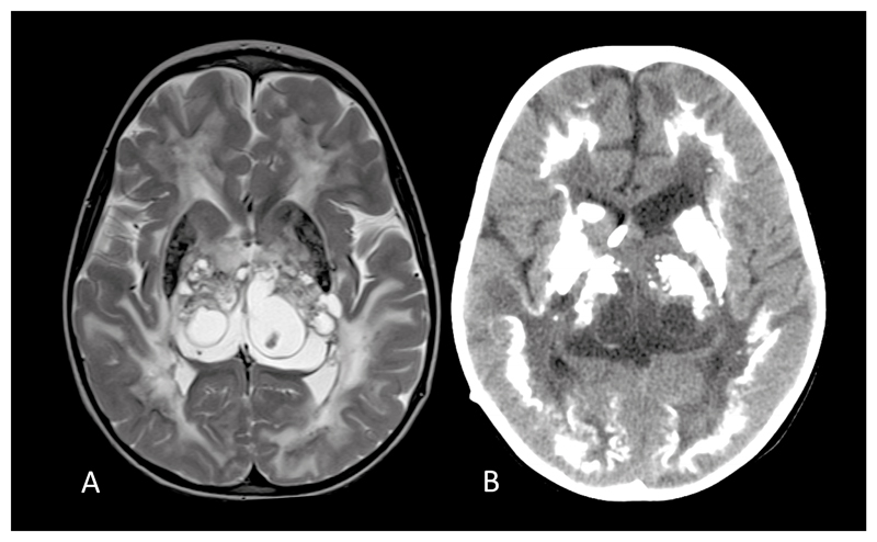
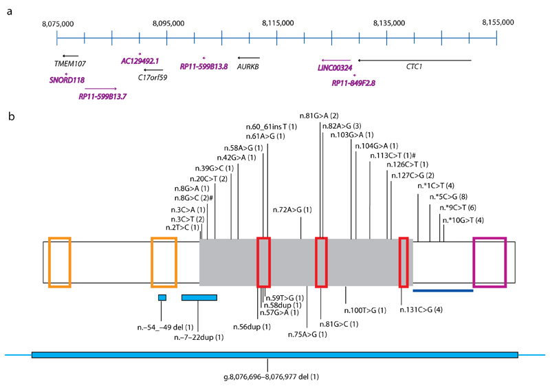
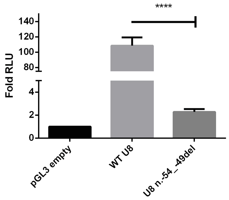
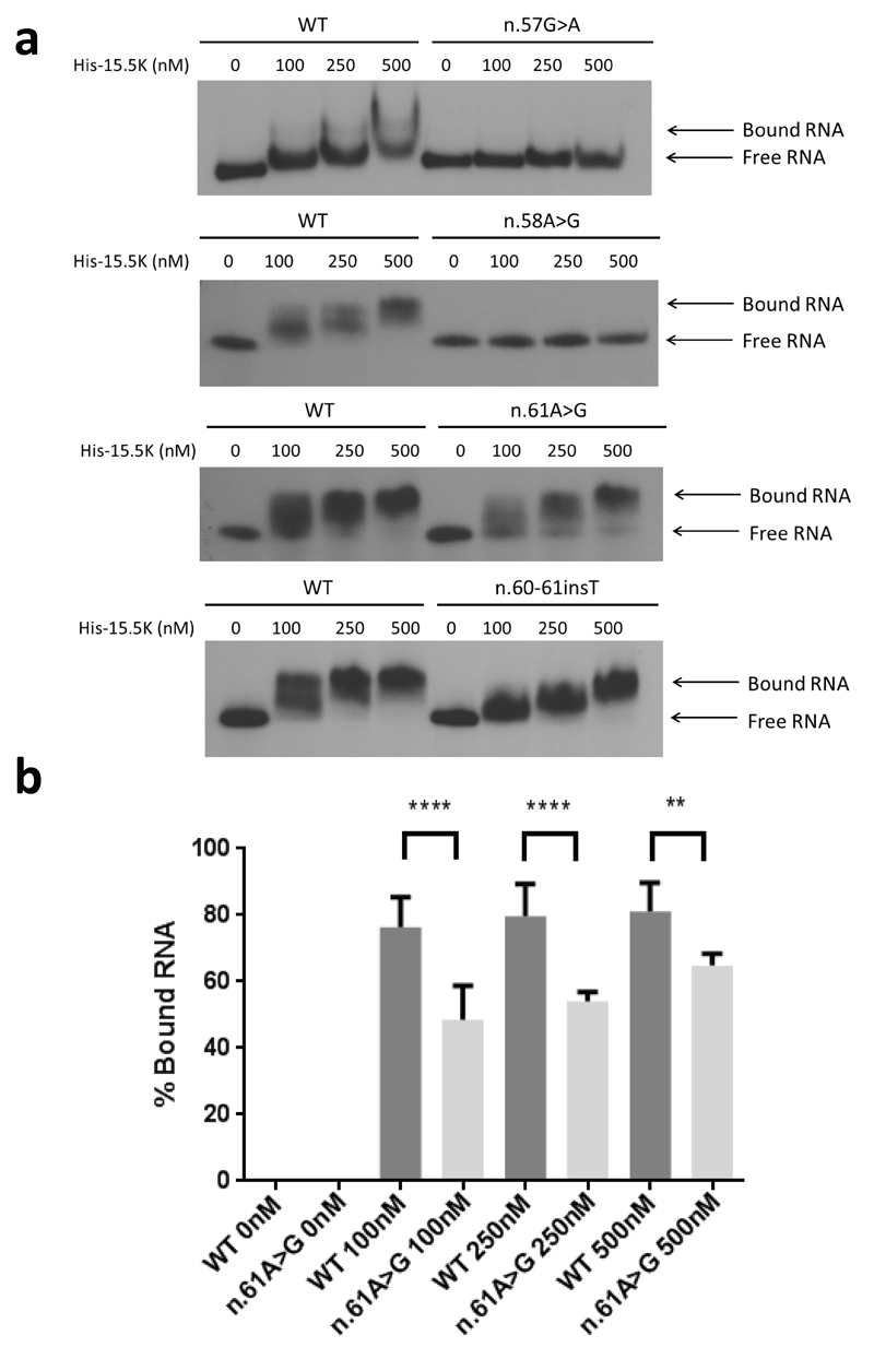
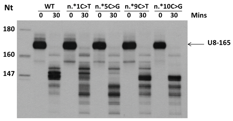
Although ribosomes are ubiquitous and essential for life, recent data indicate that monogenic causes of ribosomal dysfunction can confer a remarkable degree of specificity in terms of human disease phenotype. Box C/D small nucleolar RNAs (snoRNAs) are evolutionarily conserved non-protein-coding RNAs involved in ribosome biogenesis. Here we show that biallelic mutations in the gene SNORD118, encoding the box C/D snoRNA U8, cause the cerebral microangiopathy leukoencephalopathy with calcifications and cysts (LCC), presenting at any age from early childhood to late adulthood. These mutations affect U8 expression, processing and protein binding and thus implicate U8 as essential in cerebral vascular homeostasis.


Conflict of interest statement

The authors declare that they have no competing financial interests.

Figures

Fig. 1
Fig. 1. Typical magnetic resonance (MR) and computed tomography (CT) appearances of LCC.
(a). Axial T2 cranial MR at 4 years of age of patient F172 demonstrating symmetrical high signal of the periventricular, deep and subcortical white matter, multiple cysts within the thalami and basal ganglia, and calcification involving the putamen. (b). Cranial CT of the same patient at age 5 years showing dense, rock-like calcification in the basal ganglia and thalami. There is also dense calcification of the deep cortex and some deep white matter calcification.
Fig. 2
Fig. 2. Schematic of chromosome 17p13.1 and SNORD118.
(a) Genes across chromosome 17p13.1 between 8,075,000 and 8,155,000 are drawn to scale (numbered according to GRCh37). Protein encoding genes are represented in black text, whilst non-protein encoding genes (including SNORD118) are annotated in purple. SNORD118 lies within the 3’ UTR of TMEM107 and 50 kb from CTC1. (b) Positions of variants identified in SNORD118. Green box represents mature SNORD118. From 5’ to 3’, the orange boxes highlight the distal (DSE) and proximal (PSE) sequence elements. From 5’ to 3’, the C box, LSm and D box binding sites of SNORD118 are shown by the red boxes. The violet box represents the 3’ box (end of precursor transcript). The blue line represents the sequence encompassing the 3’ precursor transcripts of SNORD118 which are intermediates of the mature transcript. Variants that have been seen on the ExAC browser are shown above the box, with novel variants not seen on ExAC shown below. The number of LCC families with each variant is shown in brackets. Deletions and duplications are represented by blue boxes beneath the schematic. # In F344, both of these rare variants were seen in the homozygous state. However, n.8G>C was also observed in F278, suggesting that this is the likely pathogenic variant.
Fig. 3
Fig. 3. Variant n.-54_-49del found in F454 reduces the activity of the PSE element in dual luciferase assays.
HeLa cells were transfected with the Promega pGL3 reporter vector carrying the wild type PSE without the deletion (WT U8), with n.-54_-49del (U8 n.-54_-49del), or the reporter vector without an insert (pGL3 empty). The WT U8 PSE vector functioned as a promoter, enhancing luciferase activity by a mean of 109-fold in comparison with empty vector. In contrast, the n.-54_-49del vector demonstrated a mean of 2 fold activity compared to empty vector. Data presented relate to the mean fold change (+/- SD) of relative light units (RLU) compared to the control vector for three independent experiments each with three technical replicates. Data were analyzed using a one way Anova with multiple comparisons where **** = p<0.0001.
Fig. 4
Fig. 4. Protein binding of U8 variants.
(a). Electrophoretic mobility shift assay (EMSA) using wild type (WT) and mutant 5’ end-labeled in vitro transcribed U8 snoRNA with increasing concentrations of recombinant 6His-tagged 15.5K protein (His-15.5K). The concentration of the recombinant protein is given in nM above the panels. Binding of WT RNA resulted in a shift in mobility at 100 nM. Binding of His-15.5K with n.57G>A, and n.58A>G was severely impaired. A shift in mobility could not be observed for n.57G>A or n.58A>G at protein concentrations up to 500 nM. Binding between His-15.5K and n.61A>G demonstrated a shift in mobility at 100 nM; however, this shift was less than observed in WT RNA at the same concentration, and excess free RNA can be seen at all concentrations up to 500 nM indicating that binding is impaired. Similarly binding between His-15.5K and n.60_61insT demonstrated a shift in mobility at 100 nM. This shift was less than observed in WT RNA and equivalent shift was only achieved at the highest concentration tested 500nM, indicating that binding is impaired. (b). Quantification of binding between His 15.5K and n.61A>G compared to WT RNA. The percentage of protein bound RNA is significantly decreased at all concentrations. Data are given as the mean +/- SD; n= 4 independent experiments. Data were analyzed using a one way Anova with multiple comparisons where **** = p<0.0001.
Fig. 5
Fig. 5. 3’ end precursor processing of U8 variants.
In vitro 3’ end processing of 5’ end labeled in vitro transcribed precursor U8 snoRNA (U8-165) in HeLa nuclear extracts. At 30 minutes, multiple pre-U8 snoRNA processing intermediates can be seen with the wild-type (WT) pre-U8 snoRNA. Four mutants (n.*1C>T, n.*5C>G, n.*9C>T and n.*10C>G) in the pre-U8 snoRNA at 1, 5, 9 and 10 nucleotides downstream of the mature 3’ end of U8 were assessed. All four mutants exhibited a difference in processing intermediates compared to WT. Furthermore, all mutant RNAs displayed additional shorter intermediates which are not present with the WT RNA.. Data are representative of 3 independent experiments.
Fig. 6
Fig. 6. Defective proliferation of LCC fibroblasts.
(a). Quantitative reverse transcription PCR (qPCR) of SNORD118 expression in three control (CTRL1, 2, 3), four LCC (F281, F454, F691, F906), and one CP patient (F345) primary fibroblast cell lines, normalized to two housekeeping genes, RNU24 and U6. RQ is equal to 2-ΔΔCt i.e. normalized fold change relative to CTRL1. Data given as mean +/- SEM; n=3 independent experiments. Data analyzed using one way Anova with multiple comparisons **** = p<0.0001. (b). Proliferation of patient (F281, F334, F691, F906) and control (CTRL1, 2, 3) fibroblasts. The passage number of patient cells was the same or lower than controls, except for F691 which had 3 more passages. Data given as mean +/- SEM; n=2 independent experiments. (c). Percentage of beta galactosidase positive control (n=3) and LCC (n=3) fibroblasts. Red bar represents median value for each group. Mann Whitney U test **p<0.01. (d). Representative histogram of fibroblasts from one patient (F906) and one control (CTRL2). Mean of Fluorescence (MOF) assessed at 30 minutes (H0) and 2 days (H48) after carboxyfluorescein succinimidyl ester (CFSE) labeling. (e). Quantification of mean CFSE fluorescence in fibroblasts from patients (n=4) and controls (n=3). Red bar represents median value for each group. Mann Whitney U test **p<0.01; n=2 independent experiments. (f). Percentage cells in early, late and total apoptosis for four patients and three controls. Red bar represents median value for each group. No significant difference by Mann Whitney U testing; n=2 independent experiments. ●CTRL1, ■CTRL2, ▲CTRL3, ◊F281, ○F334, □F691, ▿F906.

References

    1. Labrune P, et al. Extensive brain calcifications, leukodystrophy, and formation of parenchymal cysts: a new progressive disorder due to diffuse cerebral microangiopathy. Neurology. 1996;46:1297–301. - PubMed
    1. Nagae-Poetscher LM, et al. Leukoencephalopathy, cerebral calcifications, and cysts: new observations. Neurology. 2004;62:1206–9. - PubMed
    1. Corboy JR, Gault J, Kleinschmidt-DeMasters BK. An adult case of leukoencephalopathy with intracranial calcifications and cysts. Neurology. 2006;67:1890–2. - PubMed
    1. Livingston JH, et al. Leukoencephalopathy with calcifications and cysts: a purely neurological disorder distinct from coats plus. Neuropediatrics. 2014;45:175–82. - PubMed
    1. Watkins NJ, Bohnsack MT. The box C/D and H/ACA snoRNPs: key players in the modification, processing and the dynamic folding of ribosomal RNA. Wiley Interdiscip Rev RNA. 2012;3:397–414. - PubMed

Publication types

MeSH terms

Substances