. 2016 Oct 21;11(10):2908-2914.
doi: 10.1021/acschembio.6b00624. Epub 2016 Sep 12.

Bile Acid Recognition by NAPE-PLD

Affiliations

Bile Acid Recognition by NAPE-PLD

Eleonora Margheritis et al. ACS Chem Biol. .

Abstract

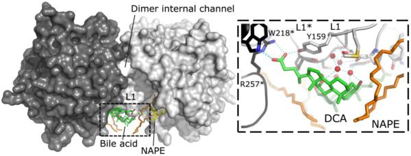
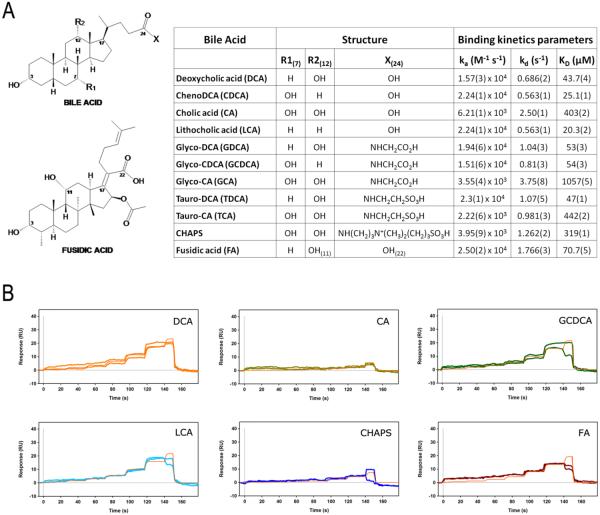
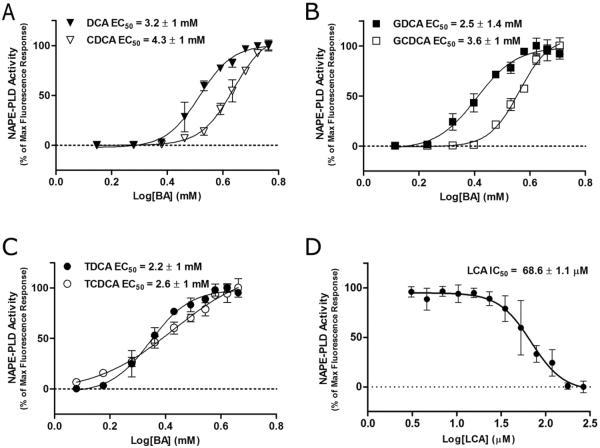
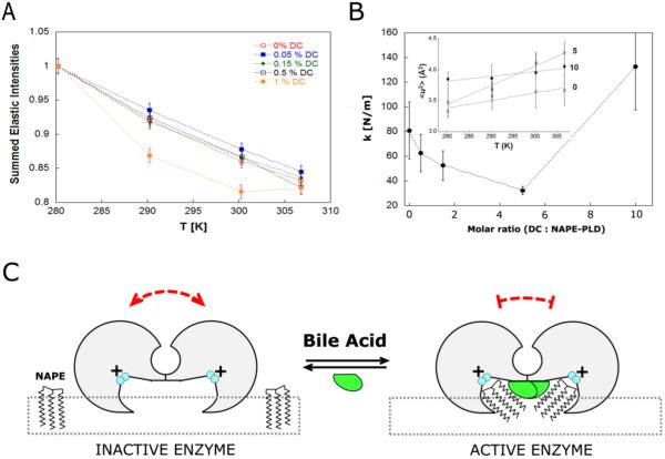
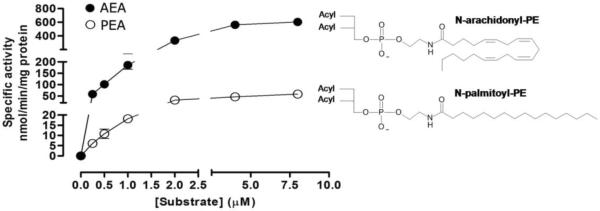
The membrane-associated enzyme NAPE-PLD (N-acyl phosphatidylethanolamine specific-phospholipase D) generates the endogenous cannabinoid arachidonylethanolamide and other lipid signaling amides, including oleoylethanolamide and palmitoylethanolamide. These bioactive molecules play important roles in several physiological pathways including stress and pain response, appetite, and lifespan. Recently, we reported the crystal structure of human NAPE-PLD and discovered specific binding sites for the bile acid deoxycholic acid. In this study, we demonstrate that in the presence of this secondary bile acid, the stiffness of the protein measured by elastic neutron scattering increases, and NAPE-PLD is ∼7 times faster to catalyze the hydrolysis of the more unsaturated substrate N-arachidonyl-phosphatidylethanolamine, compared with N-palmitoyl-phosphatidylethanolamine. Chenodeoxycholic acid and glyco- or tauro-dihydroxy conjugates can also bind to NAPE-PLD and drive its activation. The only natural monohydroxy bile acid, lithocholic acid, shows an affinity of ∼20 μM and acts instead as a reversible inhibitor (IC50 ≈ 68 μM). Overall, these findings provide important insights into the allosteric regulation of the enzyme mediated by bile acid cofactors and reveal that NAPE-PLD responds primarily to the number and position of their hydroxyl groups.


Figures

Figure 1
Figure 1
Structure of human NAPE-PLD and bile acid interactions. Surface representation of the NAPE-PLD protein dimer (PDB code: 4qn9). The two subunits (dark gray and light gray) are partly separated by an internal channel having a diameter of ~9 Å. The subunits interact mainly thought their L1 loops. These loops bind bile acid molecules (carbon atoms in green) and the glycerophospholipidic substrate N-arachydonyl-PE (carbon atoms of the sn-1 and sn-2 fatty acid chain are in orange, while those of the sn-3 in yellow). The parallel orientation of the two monomers suggests that both subunits function concurrently by recruiting NAPE substrates from the membrane. The right panel shows details of the interaction between NAPE-PLD and deoxycholic acid (DCA). The bile acid carboxyl group interacts with residue Y159 of the L1 loop, together with W218 and arginine R257 of the opposite dimer subunit (highlighted by asterisks). The steroid hydroxyls form a network of hydrogen bonds (dotted lines) that involves five L1-bridging water molecules (red spheres). Single-letter abbreviations of amino acids have been used for clarity.
Figure 2
Figure 2
Bile acid recognition by NAPE-PLD. (A) Kinetics parameters of bile acids and compound analogues (CHAPS, Fusidic acid) against NAPE-PLD, determined at 25 °C using surface plasmon resonance. (B) FastStep kinetics response profiles (and their replicates) for selected compounds. The concentration profile of the association phase was created within the biosensor SensìQ Pioneer using a twofold dilution series, and 100 μM as the highest analyte concentration. Red lines show a global fit to the response data used to extract the binding constants reported in the table (A).
Figure 3
Figure 3
Modulation of NAPE-PLD enzyme activity by different natural bile acids. (A–D) Effect of different bile acids on enzyme activity, expressed as fluorescence spectroscopic changes observed during turn-on/off assay. All graphs were obtained by fluorescence-based assay following the reaction (at 25 °C for 30 min) between the enzyme (25 nM), pre-incubated with increasing concentrations of the bile acids and the substrate PED6. LCA was tested in the presence of DCA (0.2%) to solubilize the substrate (D).
Figure 4
Figure 4
The interaction of bile acids restricts NAPE-PLD structure fluctuations. (A) Elastically scattered intensity binned over the explored range of momentum transfer (Q) as a function of temperature for NAPE-PLD at different concentrations of the bile acid DCA (left panel). (B) Dependence of the force constant k (resilience) upon the molar ratio [DCA : NAPE-PLD]. Inset: temperature dependence of the mean square displacements (< u2 >) for the complex between NAPE-PLD and DCA, at molar ratio 0, 5 and 10. (C) Hypothetical mechanism of bile acid-mediated restriction of protein dynamics. Functional, structural and dynamics measurements are consistent with a model where bile acids (green) promote the assembly of inactive NAPE-PLD subunits (gray) into an active dimer, which has a reduced dynamics. The resulting lipid-protein complex can recognize the substrate NAPE at the membrane interface (square dots) to promote its hydrolysis. The positively charged binuclear zinc center of the active site is colored in cyan.
Figure 5
Figure 5
Rate of the bile-acid activated enzyme versus NAPE. Human NAPE-PLD (0.3 μg of protein) were allowed to react with various concentrations of the substrates N-arachidonoyl-PE and N-palmitoyl-PE in the presence of the cofactor DCA (0.2%, corresponding to the maximum enzyme activation), at 37 °C for 15 min. The graph shows actual data points for the products arachidonylethanolamide (AEA, solid symbols) and palmitoylethanolamide (PEA, open symbols), their best fit line (Prism software). Km values for the two substrates were 9.6 (± 4.2) μM and 9.2 (± 1.9) μM, respectively. The maximum velocity (Vmax) resulted 1131 (± 124.4) nmol/min/mg protein for N-arachidonoyl-PE and 155.8 (± 38.36) nmol/min/mg protein for N-palmitoyl-PE. UPLC-MS/MS assays were repeated three times for each substrate.

References

    1. Wymann MP, Schneiter R. Lipid signalling in disease. Nat. Rev. Mol. Cell Biol. 2008;9:162–176. - PubMed
    1. Piomelli D. The molecular logic of endocannabinoid signalling. Nat. Rev. Neurosci. 2003;4:873–884. - PubMed
    1. Williams CM, Kirkham TC. Anandamide induces overeating: mediation by central cannabinoid (CB1) receptors. Psychopharmacology (Berl) 1999;143:315–317. - PubMed
    1. Zygmunt PM, Petersson J, Andersson DA, Chuang H, Sorgard M, Di Marzo V, Julius D, Hogestatt ED. Vanilloid receptors on sensory nerves mediate the vasodilator action of anandamide. Nature. 1999;400:452–457. - PubMed
    1. Di Marzo V, Goparaju SK, Wang L, Liu J, Batkai S, Jarai Z, Fezza F, Miura GI, Palmiter RD, Sugiura T, Kunos G. Leptin-regulated endocannabinoids are involved in maintaining food intake. Nature. 2001;410:822–825. - PubMed

Publication types