

MRI monitoring of monocytes to detect immune stimulating treatment response in brain tumor
- PMID: 27571884
- PMCID: PMC5464321
- DOI: 10.1093/neuonc/now180
MRI monitoring of monocytes to detect immune stimulating treatment response in brain tumor
Abstract
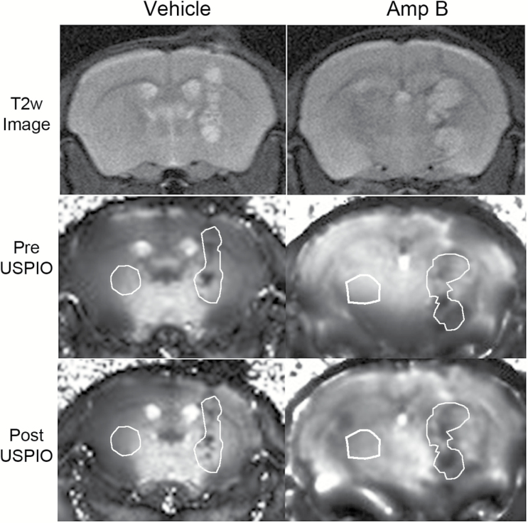
Background: Glioblastoma (GBM) is an aggressive brain cancer with a poor prognosis. The use of immune therapies to treat GBM has become a promising avenue of research. It was shown that amphotericin B (Amp B) can stimulate the innate immune system and suppress the growth of brain tumor initiating cells (BTICs). However, it is not feasible to use histopathology to determine immune activation in patients. We developed an MRI technique that can rapidly detect a therapeutic response in animals treated with drugs that stimulate innate immunity. Ultra-small iron oxide nanoparticles (USPIOs) are MRI contrast agents that have been widely used for cell tracking. We hypothesized that the increased monocyte infiltration into brain tumors due to Amp B can be detected using USPIO-MRI, providing an indicator of early drug response.
Methods: We implanted human BTICs into severe combined immunodeficient mice and allowed the tumor to establish before treating the animals with either Amp B or vehicle and then imaged them using MRI with USPIO (ferumoxytol) contrast.
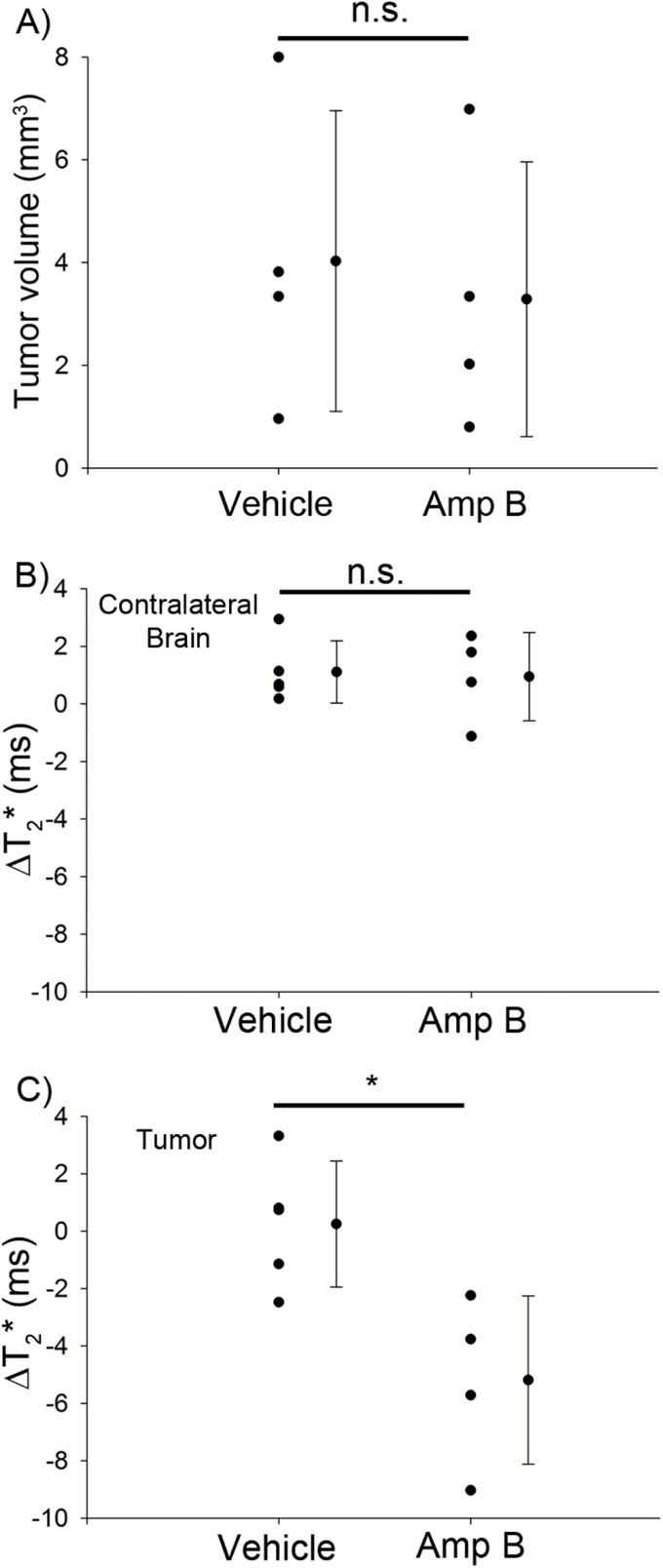
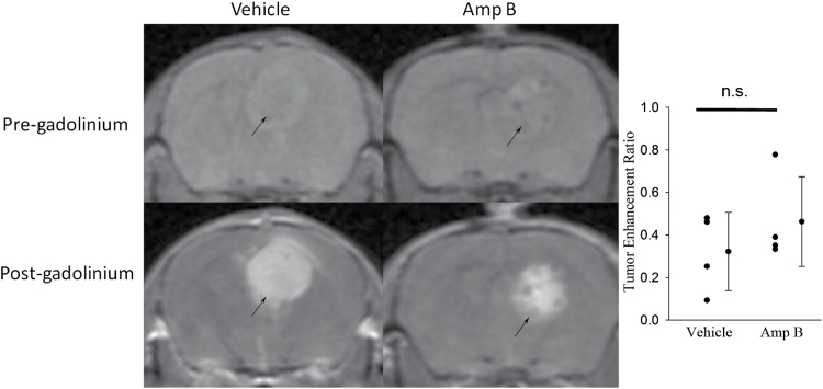
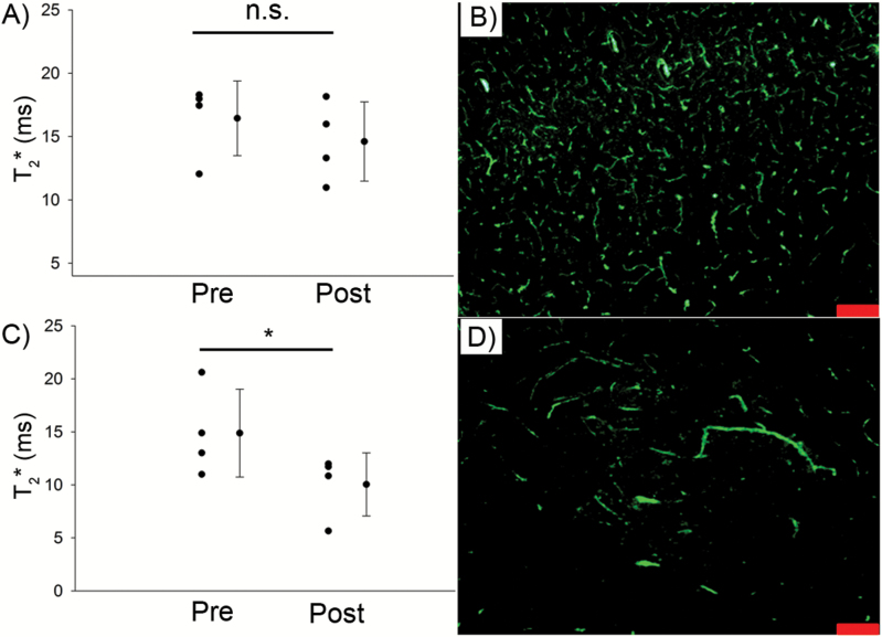
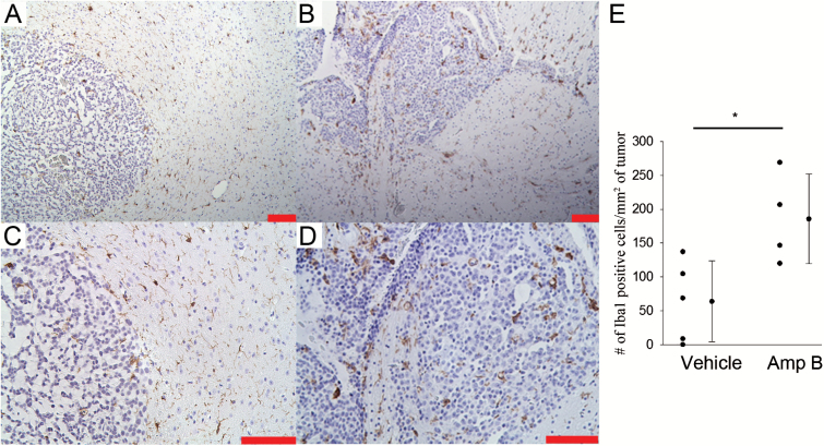
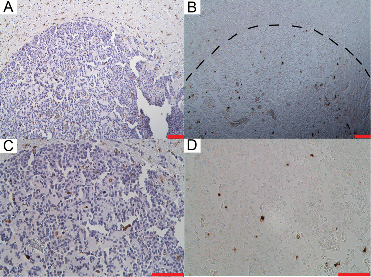
Results: After 7 days of treatment, there was a significantly decreased T2* in the tumor of Amp B but not vehicle animals, suggesting that USPIO is carried into the tumor by monocytes. We validated our MRI results with histopathology and confirmed that Amp B-treated animals had significantly higher levels of macrophage/microglia that were colocalized with iron staining in their brain tumor compared with vehicle mice.
Conclusion: USPIO-MRI is a promising method of rapidly assessing the efficacy of anticancer drugs that stimulate innate immunity.
Keywords: drug response; glioblastoma; innate immunity; iron oxide; MRI.
© The Author(s) 2017. Published by Oxford University Press on behalf of the Society for Neuro-Oncology. All rights reserved. For permissions, please e-mail: journals.permissions@oup.com
Figures
Similar articles
-
Detecting monocyte trafficking in an animal model of glioblastoma using R2* and quantitative susceptibility mapping.Cancer Immunol Immunother. 2023 Mar;72(3):733-742. doi: 10.1007/s00262-022-03297-z. Epub 2022 Oct 4. Cancer Immunol Immunother. 2023. PMID: 36194288 Free PMC article.
-
Specific detection of CD133-positive tumor cells with iron oxide nanoparticles labeling using noninvasive molecular magnetic resonance imaging.Int J Nanomedicine. 2015 Nov 11;10:6997-7018. doi: 10.2147/IJN.S86592. eCollection 2015. Int J Nanomedicine. 2015. PMID: 26635474 Free PMC article.
-
Blood-brain barrier permeability and monocyte infiltration in experimental allergic encephalomyelitis: a quantitative MRI study.Brain. 2004 Mar;127(Pt 3):616-27. doi: 10.1093/brain/awh068. Epub 2003 Dec 22. Brain. 2004. PMID: 14691063
-
USPIO-Enhanced MRI Neuroimaging: A Review.J Neuroimaging. 2016 Mar-Apr;26(2):161-8. doi: 10.1111/jon.12318. Epub 2015 Dec 3. J Neuroimaging. 2016. PMID: 26932522 Review.
-
Imaging inflammation in acute brain ischemia.Stroke. 2007 Feb;38(2 Suppl):642-5. doi: 10.1161/01.STR.0000250048.42916.ad. Stroke. 2007. PMID: 17261707 Review.
Cited by
-
The Intrinsic Biological Identities of Iron Oxide Nanoparticles and Their Coatings: Unexplored Territory for Combinatorial Therapies.Nanomaterials (Basel). 2020 Apr 27;10(5):837. doi: 10.3390/nano10050837. Nanomaterials (Basel). 2020. PMID: 32349362 Free PMC article. Review.
-
CRISPR/Cas9-mediated knockout strategies for enhancing immunotherapy in breast cancer.Naunyn Schmiedebergs Arch Pharmacol. 2024 Nov;397(11):8561-8601. doi: 10.1007/s00210-024-03208-2. Epub 2024 Jun 22. Naunyn Schmiedebergs Arch Pharmacol. 2024. PMID: 38907847 Review.
-
Immunology Meets Bioengineering: Improving the Effectiveness of Glioblastoma Immunotherapy.Cancers (Basel). 2022 Jul 29;14(15):3698. doi: 10.3390/cancers14153698. Cancers (Basel). 2022. PMID: 35954362 Free PMC article. Review.
-
Iron Oxide Nanoparticles as Theranostic Agents in Cancer Immunotherapy.Nanomaterials (Basel). 2021 Jul 29;11(8):1950. doi: 10.3390/nano11081950. Nanomaterials (Basel). 2021. PMID: 34443781 Free PMC article. Review.
-
In Vivo MR Imaging of Tumor-Associated Macrophages: The Next Frontier in Cancer Imaging.Magn Reson Insights. 2018 Apr 26;11:1178623X18771974. doi: 10.1177/1178623X18771974. eCollection 2018. Magn Reson Insights. 2018. PMID: 29780249 Free PMC article. Review.
References
-
- Stupp R, Mason WP, van den Bent MJ, et al. Radiotherapy plus concomitant and adjuvant temozolomide for glioblastoma. N Engl J Med. 2005;352(10):987–996. - PubMed
-
- Sarkar S, Doring A, Zemp FJ, et al. Therapeutic activation of macrophages and microglia to suppress brain tumor-initiating cells. Nat Neurosci. 2014; 17(1):46–55. - PubMed
-
- Wurdinger T, Deumelandt K, van der Vliet HJ, et al. Mechanisms of intimate and long-distance cross-talk between glioma and myeloid cells: how to break a vicious cycle. Biochim Biophys Acta. 2014; 1846(2):560–575. - PubMed
MeSH terms
Substances
LinkOut - more resources
Full Text Sources
Other Literature Sources
Medical
