. 2017 Feb 1;19(2):197-207.
doi: 10.1093/neuonc/now174.

Inhibition of glioblastoma tumorspheres by combined treatment with 2-deoxyglucose and metformin

Affiliations

Inhibition of glioblastoma tumorspheres by combined treatment with 2-deoxyglucose and metformin

Eui Hyun Kim et al. Neuro Oncol. .

Abstract

Background: Deprivation of tumor bioenergetics by inhibition of multiple energy pathways has been suggested as an effective therapeutic approach for various human tumors. However, this idea has not been evaluated in glioblastoma (GBM). We hypothesized that dual inhibition of glycolysis and oxidative phosphorylation could effectively suppress GBM tumorspheres (TS).

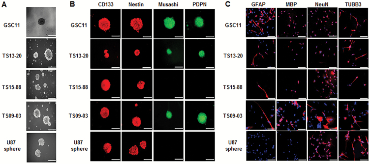
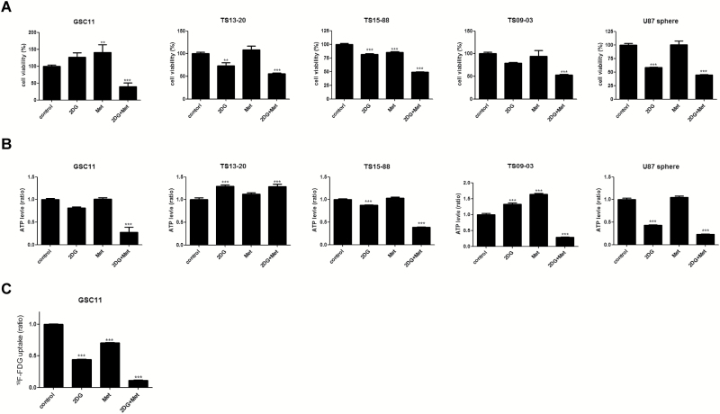
Methods: Effects of 2-deoxyglucose (2DG) and metformin, alone and in combination, on GBM-TS were evaluated. Viability, cellular energy metabolism status, stemness, invasive properties, and GBM-TS transcriptomes were examined. In vivo efficacy was tested in a mouse orthotopic xenograft model.

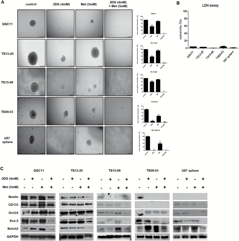
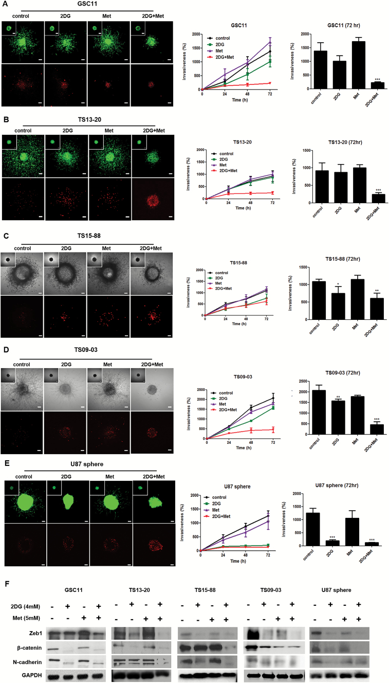
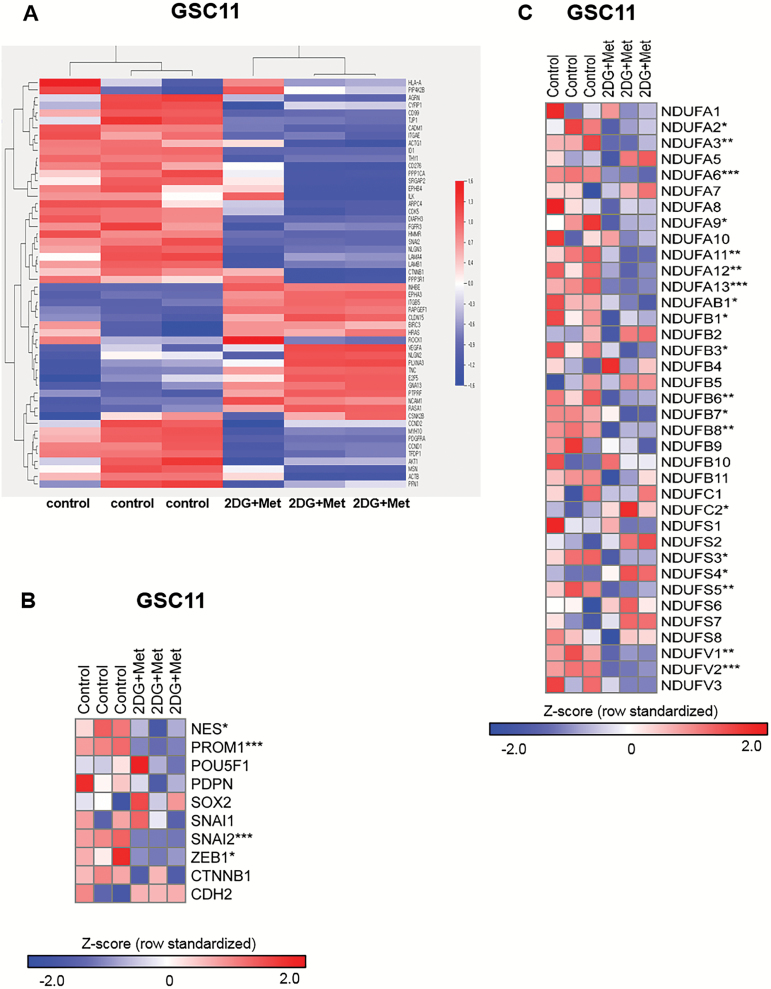
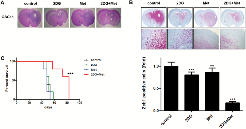
Results: GBM-TS viability was decreased by the combination of 2DG and metformin. ATP assay and PET showed that cellular energy metabolism was also decreased by this combination. Sphere formation, expression of stemness-related proteins, and invasive capacity of GBM-TS were also significantly suppressed by combined treatment with 2DG and metformin. A transcriptome analysis showed that the expression levels of stemness- and epithelial mesenchymal transition-related genes were also significantly downregulated by combination of 2DG and metformin. Combination treatment also prolonged survival of tumor-bearing mice and decreased invasiveness of GBM-TS.

Conclusion: The combination of 2DG and metformin effectively decreased the stemness and invasive properties of GBM-TS and showed a potential survival benefit in a mouse orthotopic xenograft model. Our findings suggest that targeting TS-forming cells by this dual inhibition of cellular bioenergetics warrants expedited clinical evaluation for the treatment of GBM.

Keywords: 2-deoxyglucose; glioblastoma; invasion; metformin; stemness.


Figures

Fig. 1
Fig. 1
Characterization of 5 GBM-TS. (A) TS formation was observed after 3 weeks of culture in GSC11, TS13-20, TS15-88, TS09-03, and U87 spheres. (B) Immunocytochemistry for stemness markers CD133, nestin, Musashi, PDPN. All 4 stem markers were observed in GSC11, TS13-20, and TS09-03. Musashi and PDPN were not detectable in TS15-88 and U87 spheres. (C) Neuroglial differentiation of TS. Two weeks of culture in differentiation medium resulted in successful neuroglial differentiation, as confirmed by positive GFAP, MBP, NeuN, and TUBB3 staining, although GFAP and MBP were not detectable in the U87 sphere. Nuclei were counterstained with 4′,6′-diamidino-2-phenylindole. Images are ×100 original magnification with scale bar = 50 μm for (A) and (B), ×200 original magnification with scale bar = 200 μm for (C).
Fig. 2
Fig. 2
Antiproliferative effects of 2DG and metformin and cellular energy metabolism in GBM-TS. (A) 5mM of metformin (Met) alone did not effectively inhibit proliferation of GBM-TS except TS15-88, and rather increased the viability of TS in GSC11. A moderate antiproliferative effect of 2DG (4mM) was observed only in TS13-20, TS15-88, and U87 spheres. The combination of 2DG and Met, however, constantly exhibited a significant antiproliferative effect in all 5 GBM-TS. (B) ATP assay revealed a significant decrease in ATP levels in 4 out of the 5 TS tested (no decrease observed in TS13-20) when treated with the combination of 2DG and Met. (C) 18F-FDG uptake measured by PET. In GSC11, 2DG and Met alone showed a moderate decrease in 18F-FDG uptake. When GSC11 TS were treated with these 2 simultaneously, PET indicated a much greater decrease in glucose metabolism. (**P < .01, ***P < .001, compared with the control).
Fig. 3
Fig. 3
Combined treatment with 2DG and metformin decreases stemness of GBM-TS (A) Sphere formation was tested under different conditions. 2DG (4mM) alone inhibited sphere formation with varying degrees of anti-stemness effect in all 5 TS. Treatment with 5mM of metformin (Met) alone showed less prominent anti-stemness effect compared with 2DG only in TS13-20, TS09-03, and U87 spheres. Sphere formation was nearly completely inhibited by treatment with the combination of 2DG and metformin. All images are ×50 original magnification with scale bar = 200 μm. (***P < .001, compared with the control). (B) The viability of spheres treated in different conditions was tested using the LDH assay, which revealed that the majority of sphere cells were viable, implying that decreased sphere formation was not mainly mediated by cell death. (C) Downregulation of stemness-related genes by combination treatment was confirmed by western blot analyses. Sox-2 and Notch2 were the most consistent markers when compared with the results of sphere formation assays.
Fig. 4
Fig. 4
Combined treatment with 2DG and metformin inhibits invasiveness in GBM-TS. (A), (B), (C), (D), and (E) GBM TS invasion in collagen type I matrices was significantly inhibited by combined treatment with 4mM of 2DG and 5mM of metformin (Met) in all 5 different TS. The anti-invasion effect of 2DG alone was observed in TS15-88, TS09-03, and U87 spheres. Met alone failed to inhibit invasion of GBM-TS. When compared with the control, red fluorescence indicating dead cells was a little increased in 2DG, Met, and their combination treatment groups; however, the majority of red fluorescing cells were localized in the core of spheroids. Images were obtained after 72 hours. Scale bar: 100 μm. (*P < .05, **P < .01, ***P < .001, compared with the control at 72 hours). (F) Western blotting of EMT-related genes; Zeb1, β-catenin, and N-cadherin. Decreased expression of EMT-related genes was observed in all TS treated by combination of 2DG and Met. The downregulation of EMT-related gene expression by 2DG treatment alone was mostly observed in all TS.
Fig. 5
Fig. 5
Gene expression microarray and class comparison. (A) Heatmap of genes differentially expressed before and after 2DG/metformin (Met)-treatment. (B) Genes of interest. Combined treatments of 2DG and Met downregulated gene expression of stemness- and EMT-related genes; NES (nestin), PROM1 (CD133), SNAI2 (TWIST), and ZEB1. (C) Expression levels of mitochondrial complex I genes were evaluated using microarray experiment. The values were z-transformed using whole genes (n = 3, *P < .05, **P < .01, ***P < .001 by Student’s t-test).
Fig. 6
Fig. 6
Effects of combined treatment with 2DG and metformin in an orthotopic xenograft model. Sections of mouse brains, obtained from euthanized mice at the end of the experiment, were H&E stained to show the margins of gross tumors (A), and immunostained for Zeb1 to identify invading cells (B). Original magnification, ×12. Combination treatment of 2DG and metformin (Met) markedly inhibited invasion of GBM-TS compared with the control and other single treatments. (C) Kaplan‒Meier survival curve showed increased survival of mice treated with the combination of 2DG and Met compared with the control other single treatments. (***P < .001).

Similar articles

Cited by

References

    1. Bai RY, Staedtke V, Riggins GJ. Molecular targeting of glioblastoma: drug discovery and therapies. Trends Mol Med. 2011;17(6):301–312. - PMC - PubMed
    1. Stupp R, Hegi ME, Mason WP, et al. Effects of radiotherapy with concomitant and adjuvant temozolomide versus radiotherapy alone on survival in glioblastoma in a randomised phase III study: 5-year analysis of the EORTC-NCIC trial. Lancet Oncol. 2009;10(5):459–466. - PubMed
    1. Liu G, Yuan X, Zeng Z, et al. Analysis of gene expression and chemoresistance of CD133+ cancer stem cells in glioblastoma. Mol Cancer. 2006;5:67. - PMC - PubMed
    1. Bao S, Wu Q, McLendon RE, et al. Glioma stem cells promote radioresistance by preferential activation of the DNA damage response. Nature. 2006;444(7120):756–760. - PubMed
    1. Binda E, Reynolds BA, Vescovi AL. Glioma stem cells: turpis omen in nomen? (The evil in the name?). J Intern Med. 2014;276(1):25–40. - PubMed

MeSH terms