. 2016 Aug 30:6:32351.
doi: 10.1038/srep32351.

Forced co-expression of IL-21 and IL-7 in whole-cell cancer vaccines promotes antitumor immunity

Affiliations

Forced co-expression of IL-21 and IL-7 in whole-cell cancer vaccines promotes antitumor immunity

Yang-Zhuo Gu et al. Sci Rep. .

Abstract

Genetic modification of whole-cell cancer vaccines to augment their efficacies has a history of over two and a half decades. Various genes and gene combinations, targeting different aspects of immune responses have been tested in pursuit of potent adjuvant effects. Here we show that co-expression of two cytokine members of the common cytokine receptor γ-chain family, IL-21 and IL-7, in whole-cell cancer vaccines boosts antitumor immunity in a CD4(+) and CD8(+) T cell-dependent fashion. It also generates effective immune memory. The vaccine-elicited short-term effects positively correlated with enhanced infiltration of CD4(+) and CD8(+) effector T cells, and the long-term effects positively correlated with enhanced infiltration of effector memory T cells, especially CD8(+) effector memory T cells. Preliminary data suggested that the vaccine exhibited good safety profile in murine models. Taken together, the combination of IL-21 and IL-7 possesses potent adjuvant efficacy in whole-cell vaccines. This finding warrants future development of IL-21 and IL-7 co-expressing whole-cell cancer vaccines and their relevant combinatorial regimens.


Figures

Figure 1
Figure 1. Establishment of vaccine cell lines by lentiviral transduction.
(A) Lentiviral constructs used in this study. LTR, long terminal repeat. Ψ, packaging signal. RRE, Rev response element. cPPT, central polypurine tract. CMVp, CMV promoter. IRES, internal ribosome entry site. Bsd, blasticidin resistance gene. WPRE, woodchuck hepatitis virus posttranscriptional regulatory element. Sp, Signal peptide of IL-7. FA, furin cleavage site and P2A peptide. (B) Western blot analysis of secreted IL-21 and IL-7 in vaccine cell-conditioned media. Note that the two bands of IL-21 in the 21/7 CM lane represented two possible cleavage products: cleavage at furin site resulted in IL-21 + 4AA, while cleavage at P2A site resulted in IL-21 + 25AA. CM, conditioned medium. AA, amino acids. (C) Western blot analysis of STAT proteins activated in response to secreted IL-21 and IL-7. SC, splenocytes. MC, medium control. Full-length blots are presented in Supplementary Figure S1.
Figure 2
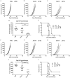
Figure 2. Vaccination with IL-21 and IL-7 co-expressing B16F10 cells prevented tumorigenesis of B16F10 melanoma in prophylactic setting.
(A–E) Individual tumor growth curves of B16F10 tumors. Mice were vaccinated s.c. on day −14 and day −7 with 106 irradiated tumor cells, then challenged c.l. on day 0 with 105 viable B16F10 cells. Fractions in the parentheses indicate the proportion of tumor-free mice in totals. Data were pooled from two independent repeats. (F) Cumulative survival curves of two independent repeats. **P < 0.01. ****P < 0.0001. (G–J) Individual tumor growth curves of B16F10 tumors. Mice were vaccinated s.c. on day −14 and day −7 with PBS or 106 irradiated tumor cells following one of the following regimens. 1) prime with 16–21 cells, boost with 16-7 cells in (H). 2) prime with 16-7 cells, boost with 16–21 cells in (I). 3) prime and boost with a mixture of 5 × 105 16–21 and 5 × 105 16-7 cells in (J). mice were then challenged c.l. on day 0 with 105 viable B16F10 cells. Fractions in the parentheses indicate the proportion of tumor-free mice in totals. Data were pooled from two independent repeats. (K) Cumulative survival curves of two independent repeats.
Figure 3
Figure 3. Vaccination with IL-21 and IL-7 co-expressing cells inhibited tumor progression in two therapeutic models.
(A–C) Individual tumor growth curves of B16F10 tumors. Mice were challenged s.c. on day 0 with 5 × 104 viable B16F10 cells, then vaccinated c.l. on day 3 with two doses of 107 irradiated tumor cells. Data were pooled from two independent repeats. (D) Cumulative tumor volumes on day 19 of two independent repeats. Horizontal bars represent means. (E) Cumulative survival curves of two independent repeats. (F–H) Individual tumor growth curves of CT26 tumors. Mice were challenged s.c. on day 0 with 105 viable CT26 cells, then vaccinated c.l. on day 3 with two doses of 107 irradiated tumor cells. Fractions in the parentheses indicate the proportion of cured mice in totals. Data were pooled from two independent repeats. (I) Cumulative tumor volumes on day 19 of two independent repeats. Horizontal bars represent means. (J) Cumulative survival curves of two independent repeats. ***P < 0.001. ****P < 0.0001.
Figure 4
Figure 4. Antitumor efficacies of IL-21 and IL-7 co-expressing tumor cell vaccine depended highly on CD4+ and CD8+ T cells, rather than NK cells.
(A–F) Individual tumor growth curves of B16F10 tumors. Wild-type, as well as CD4 KO and CD8 KO mice were vaccinated s.c. on day −14 and day −7 with 106 irradiated tumor cells, then challenged c.l. on day 0 with 105 viable B16F10 cells. Fractions in the parentheses indicate the proportion of tumor-free mice in totals. Data were pooled from two independent repeats. (G) Cumulative survival curves of two independent repeats. ***P < 0.001. ****P < 0.0001. (H–K) Individual tumor growth curves of B16F10 tumors. Mice were vaccinated s.c. on day −14 and day −7 with 106 irradiated tumor cells, then challenged c.l. on day 0 with 105 viable B16F10 cells. PBS, isotype control, or NK1.1 antibody were administrated i.p. following the protocol described in Methods. Fractions in the parentheses indicate the proportion of tumor-free mice in totals. Data were pooled from two independent repeats. (L) Cumulative survival curves of two independent repeats.
Figure 5
Figure 5. Vaccination with IL-21 and IL-7 co-expressing tumor cells enhanced CD8+ and CD4+ effector T cell infiltration in tumors and raised infiltrating effector/regulatory cell ratios.
(A–C) Frequencies of CD8+, CD4+ effector and regulatory cells in total cells of B16F10 tumors generated by challenge s.c. with 3 × 106 viable cells at day 0. Data were pooled from two independent repeats. (D,E) CD8+/Treg and CD4+ Teff/Treg ratios in B16F10 tumors. Data were pooled from two independent repeats. Horizontal bars represent means. *P < 0.05. **P < 0.01.
Figure 6
Figure 6. Vaccination with IL-21 and IL-7 co-expressing B16F10 cells elicited memory responses.
(A,B) Individual tumor growth curves of B16F10 tumors. Mice remaining tumor-free were rechallenged with 2 × 105 viable B16F10 cells on day 90. Fractions in the parentheses indicate the proportion of tumor-free mice in totals. Data were pooled from two independent repeats. (C) Cumulative survival curves of two independent repeats. ****P < 0.0001. (D,E) Frequencies of CD8+, CD4+ effector memory T cells in total cells of B16F10 tumors generated by challenge with 4 × 105 viable cells at day 35. Data were pooled from two independent repeats. Horizontal bars represent means. *P < 0.05. **P < 0.01.
Figure 7
Figure 7. Vaccination with IL-21 and IL-7 co-expressing tumor cells exhibited good safety profile with trivial side effect.
(A) Vaccination with IL-21 and IL-7 co-expressing tumor cells induced depigmentation. Upper arrows in (a) and (b) indicate vitiligo at challenge sites. Lower arrow in (b) indicates vitiligo at boost site. (B) H&E staining of vital organs of vaccinated mice. Scale bar, 100 μm.

Similar articles

Cited by

References

    1. Tepper R. I., Pattengale P. K. & Leder P. Murine interleukin-4 displays potent anti-tumor activity in vivo. Cell 57, 503–512 (1989). - PubMed
    1. Watanabe Y. et al.. Exogenous expression of mouse interferon gamma cDNA in mouse neuroblastoma C1300 cells results in reduced tumorigenicity by augmented anti-tumor immunity. Proc Natl Acad Sci USA 86, 9456–9460 (1989). - PMC - PubMed
    1. Dranoff G. et al.. Vaccination with irradiated tumor cells engineered to secrete murine granulocyte-macrophage colony-stimulating factor stimulates potent, specific, and long-lasting anti-tumor immunity. Proc Natl Acad Sci USA 90, 3539–3543 (1993). - PMC - PubMed
    1. Leonard W. J. & Spolski R. Interleukin-21: a modulator of lymphoid proliferation, apoptosis and differentiation. Nat Rev Immunol 5, 688–698 (2005). - PubMed
    1. Rochman Y., Spolski R. & Leonard W. J. New insights into the regulation of T cells by gamma(c) family cytokines. Nat Rev Immunol 9, 480–490 (2009). - PMC - PubMed

Publication types

MeSH terms