. 2016 Jan 27:1:15021.
doi: 10.1038/nmicrobiol.2015.21.

The microenvironment of injured murine gut elicits a local pro-restitutive microbiota

Affiliations

The microenvironment of injured murine gut elicits a local pro-restitutive microbiota

Ashfaqul Alam et al. Nat Microbiol. .

Abstract

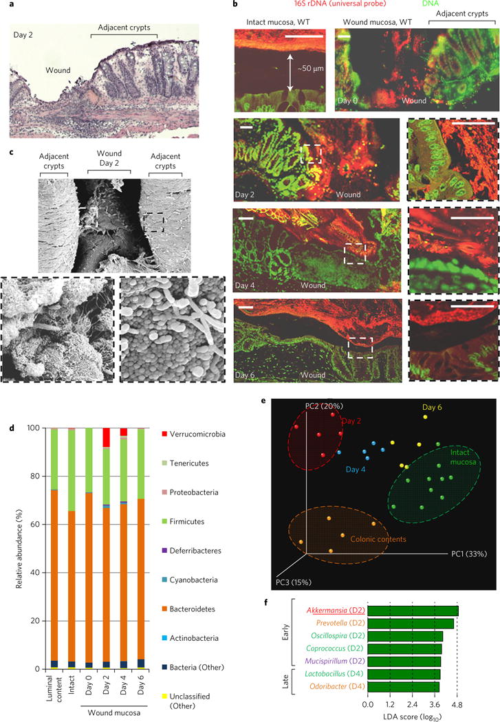
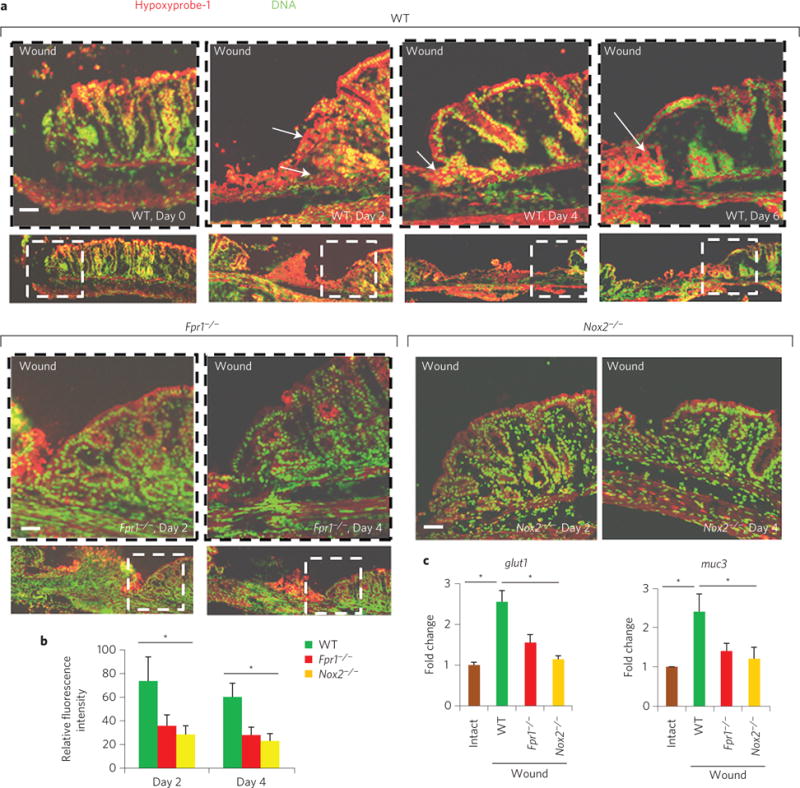
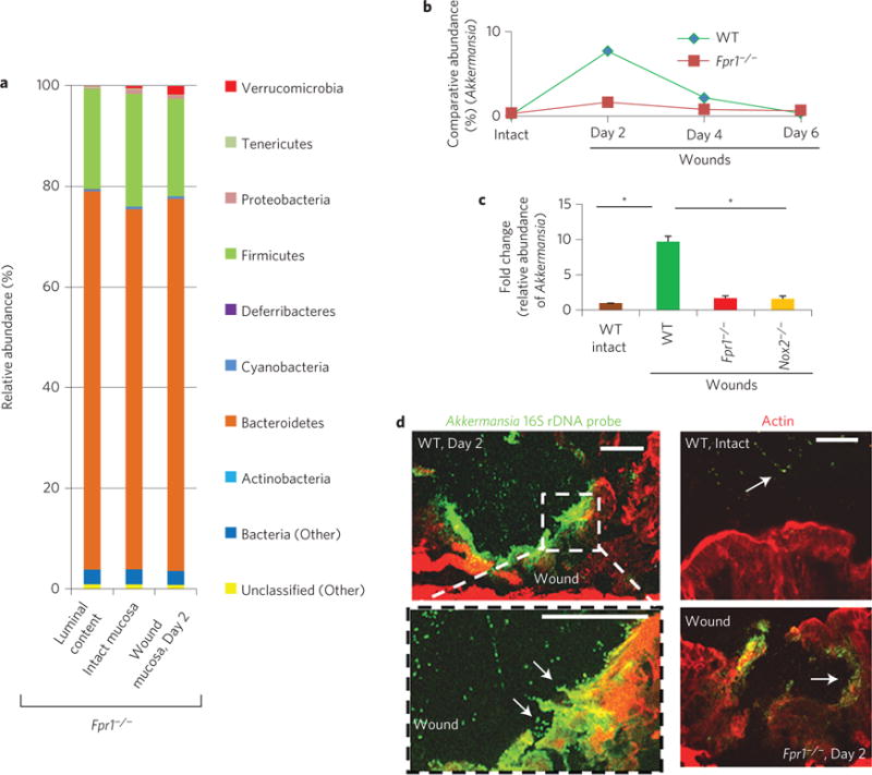
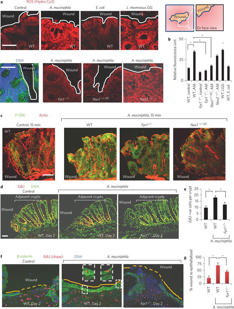
The mammalian intestine houses a complex microbial community, which influences normal epithelial growth and development, and is integral to the repair of damaged intestinal mucosa(1-3). Restitution of injured mucosa involves the recruitment of immune cells, epithelial migration and proliferation(4,5). Although microenvironmental alterations have been described in wound healing(6), a role for extrinsic influences, such as members of the microbiota, has not been reported. Here, we show that a distinct subpopulation of the normal mucosal-associated gut microbiota expands and preferentially colonizes sites of damaged murine mucosa in response to local environmental cues. Our results demonstrate that formyl peptide receptor 1 (FPR1) and neutrophilic NADPH oxidase (NOX2) are required for the rapid depletion of microenvironmental oxygen and compensatory responses, resulting in a dramatic enrichment of an anaerobic bacterial consortium. Furthermore, the dominant member of this wound-mucosa-associated microbiota, Akkermansia muciniphila (an anaerobic, mucinophilic gut symbiont(7,8)), stimulated proliferation and migration of enterocytes adjacent to the colonic wounds in a process involving FPR1 and intestinal epithelial-cell-specific NOX1-dependent redox signalling. These findings thus demonstrate how wound microenvironments induce the rapid emergence of 'probiont' species that contribute to enhanced repair of mucosal wounds. Such microorganisms could be exploited as potential therapeutics.


Conflict of interest statement

The authors declare no competing financial interests.

Figures

Figure 1
Figure 1. Restitutive wound induces spatiotemporal change of wound mucosa-associated microbiota
a, H&E-stained wound bed. b, FISH of mucosal wound beds (n = 10 per group) using a pan-bacterial probe (red). Scale bars, 50 μm. c, Scanning electron micrograph of wound bed. d, Mean relative abundance of bacterial phyla determined by HTS (n = 5 wounds per group). e, PCoA plot of microbiota community structure in injured mucosa. f, LDA scores reflecting positive association of bacterial genus with restitutive mucosa. D2 and D4 denote day 2 and day 4, respectively.
Figure 2
Figure 2. FPR1/NOX2 is required for microenvironmental changes in restitutive wounds, which promote anaerobic mucinophilic Akkermansia
a, Immunofluorescence staining of pimonidazole HCl showing depletion of microenvironmental oxygen in mucosal wound beds (n = 10 per group). Arrows indicate areas of hypoxia. b, Quantitative representation of immunofluorescence analysis (by ImageJ software; expressed in units of fluorescence; mean ± s.e.m.) of pimonidazole HCl adduct staining in a. c, Real-time qPCR analysis (mean ± s.e.m.) of mucosal epithelial hypoxia response genes glut1 and muc3 in wounds (n = 11 per group). Scale bars, 50 μm. *P < 0.05; Student’s t-test.
Figure 3
Figure 3. FPR1 is required for enrichment of Akkermansia sp
a, Relative abundance of bacterial phyla determined by HTS from Fpr1/ mice. Data represent mean of relative abundance with n = 5 wounds per group. b, Comparative relative abundance of Akkermansia in wounds in WT and Fpr1/ mice determined by HTS. n = 5 wounds/group. c, Real-time qPCR analysis of fold change in specific relative abundance of Akkermansia (mean ± s.e.m; n = 10 per group). d, Visualization of Akkermansia (green) in wound beds (n = six per group) by FISH. Scale bars, 50 μm. *P < 0.05; Student’s t-test.
Figure 4
Figure 4. Akkermansia muciniphila enhances redox-dependent wound restitution
a, Cellular ROS generation in mucosal wounds oriented en face (n = 10 per group) after luminal treatments of wounds as in ref. . DNA, blue; actin, green. b, Quantitative representation of ROS production (mean ± s.e.m) in a. c, Immunofluorescence analysis of P-ERK in thin sections of colonic wounds (n = 11 per group). d, Enterocyte proliferation (EdU-positive cells in red) after intrarectal administration of A. muciniphila. e, Quantification of EdU-positive cells in five crypts adjacent to wounds (n = 12 per group). f, Re-epithelialization of wound beds (n = 12 per group) with EdU-labelled (red, chase assay) enterocytes in WT and Fpr1/ mice. g, Quantification of wound re-epithelialization (mean ± s.e.m.) in f. Scale bars, 50 μm; *P < 0.05; Student’s t-test.

Similar articles

Cited by

References

    1. Hooper LV, et al. Molecular analysis of commensal host-microbial relationships in the intestine. Science. 2001;291:881–884. - PubMed
    1. Backhed F, Ley RE, Sonnenburg JL, Peterson DA, Gordon JI. Host–bacterial mutualism in the human intestine. Science. 2005;307:1915–1920. - PubMed
    1. Pull SL, Doherty JM, Mills JC, Gordon JI, Stappenbeck TS. Activated macrophages are an adaptive element of the colonic epithelial progenitor niche necessary for regenerative responses to injury. Proc Natl Acad Sci USA. 2005;102:99–104. - PMC - PubMed
    1. Lotz MM, et al. Intestinal epithelial restitution. Involvement of specific laminin isoforms and integrin laminin receptors in wound closure of a transformed model epithelium. Am J Pathol. 1997;150:747–760. - PMC - PubMed
    1. Miyoshi H, Ajima R, Luo CT, Yamaguchi TP, Stappenbeck TS. Wnt5a potentiates TGFβ signaling to promote colonic crypt regeneration after tissue injury. Science. 2012;338:108–113. - PMC - PubMed