. 2018 Jan;38(1):87-92.
doi: 10.1111/cpf.12387. Epub 2016 Aug 30.

Impact of abnormal cerebrovascular reactivity on BOLD fMRI: a preliminary investigation of moyamoya disease

Affiliations

Impact of abnormal cerebrovascular reactivity on BOLD fMRI: a preliminary investigation of moyamoya disease

Erin L Mazerolle et al. Clin Physiol Funct Imaging. 2018 Jan.

Abstract

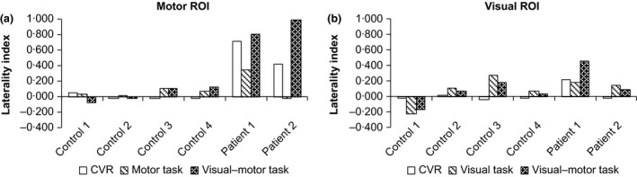
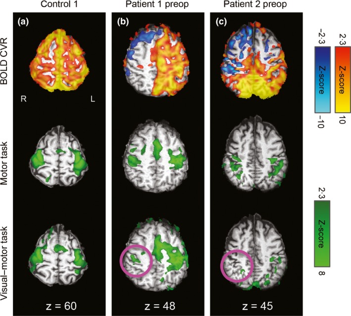
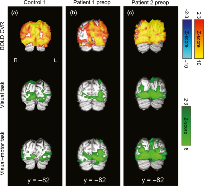
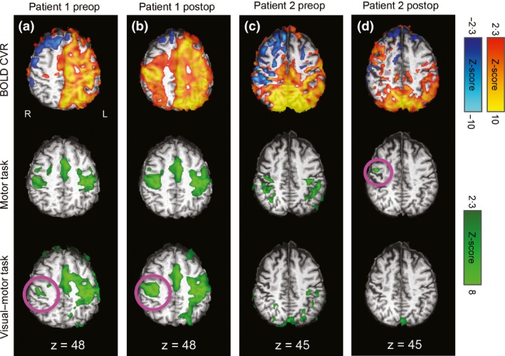
Blood oxygen level-dependent (BOLD) functional magnetic resonance imaging (fMRI) studies of patients with cerebrovascular disease have largely ignored the confounds associated with abnormal cerebral blood flow, vascular reactivity and neurovascular coupling. We studied BOLD fMRI activation and cerebrovascular reactivity in moyamoya disease. To characterize the impact of remote vascular demands on BOLD fMRI measurements, we varied the vascular territories engaged by manipulating the experimental task performed by the participants. Vascular territories affected by disease were identified using BOLD cerebrovascular reactivity. Preliminary evidence from two patients pre- and postrevascularization surgery and four controls indicates that neurovascular coupling in affected brain regions can be modulated by the task-related vascular demands in unaffected regions. In one patient studied, we observed that brain regions with improved cerebrovascular reactivity after surgery demonstrated normalized neurovascular coupling, that is the degree to which neurovascular coupling was modulated by task-related vascular demands was decreased. We propose that variations in task-dependent neurovascular coupling in patients with moyamoya disease are likely related to vascular steal. While preliminary, our findings are a proof of concept of the limitations of BOLD fMRI in cerebrovascular disease and suggest a role for assessment of cerebrovascular reactivity to improve interpretation of task-related BOLD fMRI activation.

Keywords: cerebrovascular disease; functional brain mapping; hypercapnia; neurovascular coupling; stenosis.


Figures

Figure 1
Figure 1
Laterality indices (1 = completely left‐lateralized activation; 0 = bilateral activation; −1 = completely right lateralized activation) for the four controls and two patients (preoperative data only), calculated based on the activation extent (i.e. number of significantly activated voxels). Only positive responses are considered here. a: Motor ROI; b: Visual ROI. The controls had approximately bilateral activation in both ROIs for both the separate and combined tasks, consistent with bilaterally distributed CVR. In patient 1, the motor ROI CVR was left‐lateralized. In addition, activation was left‐lateralized in both tasks, but to a greater extent in the combined task. This pattern was also observed to a lesser extent in the visual ROI in this patient. In patient 2, the motor ROI had left‐lateralized CVR, bilateral activation in the separate motor task and left‐lateralized activation in the combined visual–motor task. CVR in the visual ROI was normal in this patient, consistent with approximately bilateral activation in both the separate visual and combined visual–motor task).
Figure 2
Figure 2
CVR and task‐related activation maps overlaid on the individuals' T1‐weighted images in standard space for sensorimotor regions. R=right, L=left, coordinates refer to MNI space. Motor cortex CVR (top row) and task‐related activation during the separate motor task (middle row) and combined visual–motor task (bottom row) for an illustrative control (a), patient 1 preoperatively (b), and patient 2 preoperatively (c). Robust positive CVR was observed in the control, whereas the patients had large areas of negative or absent CVR, particularly over the affected (right) hemispheres. As expected, task activation was bilateral in the control participant (a). In the patients (b and c), activation was more lateralized during the combined task than the separate task, with reduced or absent activation over the affected hemisphere during the combined task (pink circles).
Figure 3
Figure 3
CVR and task‐related activation maps overlaid on the individuals' T1‐weighted images in standard space for visual regions. R=right, L=left, coordinates refer to MNI space. Visual cortex CVR was robustly positive in the illustrative control (a) and patient 1 (b), who both demonstrated bilateral visual activation for both the separate visual task and combined visual–motor task. There were some posterior areas with reduced/absent CVR in patient 2 (c), which corresponded to more lateralized responses, particularly in the visual–motor task, for this patient and ROI (see text and Fig. 1 for details).
Figure 4
Figure 4
Postoperative results from sensorimotor regions for patients 1 and 2 (panels b and d), with preoperative data repeated here for ease of comparison (panels a and c). R=right, L=left, coordinates refer to MNI space. Postoperatively, patient 1's activation pattern was more bilateral in both tasks, but this was particularly true for the combined task. This was consistent with improved CVR over the affected hemisphere (panel B). Patient 2's CVR was reduced postoperatively, except in a right anterior region, which was also associated with activation in the motor task (panel D, pink circle).

Similar articles

Cited by

References

    1. Andersson JL, Jenkinson M, Smith S, et al Non‐linear registration, aka Spatial normalisation FMRIB technical report TR07JA2. FMRIB Anal Group Univ Oxf (2007); Available at: http://fmrib.medsci.ox.ac.uk/analysis/techrep/tr07ja2/tr07ja2.pdf [Accessed August 5, 2015].
    1. Arteaga DF, Strother MK, Faraco CC, et al The vascular steal phenomenon is an incomplete contributor to negative cerebrovascular reactivity in patients with symptomatic intracranial stenosis. J Cereb Blood Flow Metab (2014); 34: 1453–1462. - PMC - PubMed
    1. Blair GW, Doubal FN, Thrippleton MJ, et al Magnetic resonance imaging for assessment of cerebrovascular reactivity in cerebral small vessel disease: a systematic review. J Cereb Blood Flow Metab (2016); 36: 833–841. - PMC - PubMed
    1. Burke GM, Burke AM, Sherma AK, et al Moyamoya disease: a summary. Neurosurg Focus (2009); 26: E11. - PubMed
    1. Calautti C, Baron J‐C. Functional neuroimaging studies of motor recovery after stroke in adults: a review. Stroke (2003); 34: 1553–1566. - PubMed

MeSH terms