Clinical Trial
. 2016 Aug 30:6:31531.
doi: 10.1038/srep31531.

Mapping rare, deleterious mutations in Factor H: Association with early onset, drusen burden, and lower antigenic levels in familial AMD

Affiliations
Clinical Trial

Mapping rare, deleterious mutations in Factor H: Association with early onset, drusen burden, and lower antigenic levels in familial AMD

Erin K Wagner et al. Sci Rep. .

Abstract

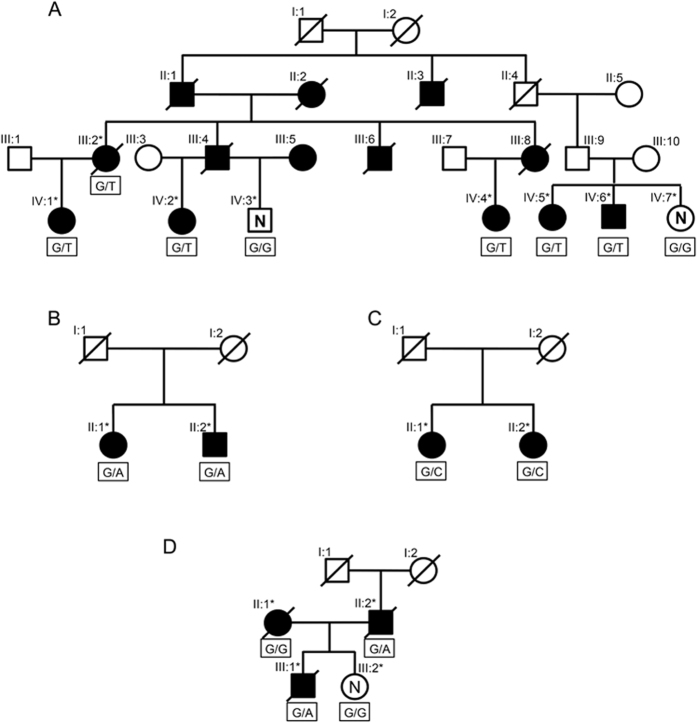
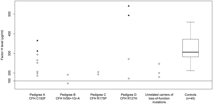
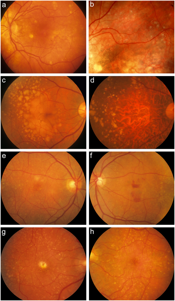
The genetic architecture of age-related macular degeneration (AMD) involves numerous genetic variants, both common and rare, in the coding region of complement factor H (CFH). While these variants explain high disease burden in some families, they fail to explain the pathology in all. We selected families whose AMD was unexplained by known variants and performed whole exome sequencing to probe for other rare, highly penetrant variants. We identified four rare loss-of-function variants in CFH associated with AMD. Missense variant CFH 1:196646753 (C192F) segregated perfectly within a family characterized by advanced AMD and drusen temporal to the macula. Two families, each comprising a pair of affected siblings with extensive extramacular drusen, carried essential splice site variant CFH 1:196648924 (IVS6+1G>A) or missense variant rs139360826 (R175P). In a fourth family, missense variant rs121913058 (R127H) was associated with AMD. Most carriers had early onset bilateral advanced AMD and extramacular drusen. Carriers tended to have low serum Factor H levels, especially carriers of the splice variant. One missense variant (R127H) has been previously shown not to be secreted. The two other missense variants were produced recombinantly: compared to wild type, one (R175P) had no functional activity and the other (C192F) had decreased secretion.


Figures

Figure 1
Figure 1. Pedigree diagrams for families carrying rare, loss-of-function CFH variants.
(A) Pedigree A (CFH C192F), (B) Pedigree B (CFH IVS6 + 1G > A), (C) Pedigree C (CFH R175P), (D) Pedigree D (CFH R127H); ⚪ = female; ◽ = male; * = sequenced; ∅ = deceased; ⦁ and ◾ = affected with advanced AMD; “N” = confirmed unaffected; empty ⚪ and ◽  = unknown affection status; rare variant genotype listed below each sequenced subject.
Figure 2
Figure 2. Serum factor H levels according to carrier state of rare CFH variants.
⚪ = subjects carrying rare CFH variant; ▴ = subjects not carrying rare CFH variant. controls = CARMS grade 1 and no known rare variants in CFH; unrelated carriers of loss-of-function mutations = nonsense, splice-site, and loss of a conserved cysteine; normal clinical laboratory range = 160–412 μg/ml.
Figure 3
Figure 3. Fundus photographs from family members carrying rare CFH variants showing numerous large drusen and extramacular drusen.
Fundus color photographs of subjects in the four pedigrees: IV:5 from Pedigree A showing several large macular and extramacular drusen with retinal pigment epithelial irregularities in her left eye (OS) at age 71 (a) and extramacular drusen and macular pigment disruption following intravitreal anti-vascular endothelial growth factor injections OS after 8 years of follow-up (b). II:1 from Pedigree B showing numerous large macular and extramacular drusen with transition in her right eye (OD) from drusenoid retinal pigment epithelial detachments at age 64 (c) to geographic atrophy at age 76 (d). Subject II:2 from Pedigree C showing several drusen throughout the posterior pole OD (e) and OS (f) at age 55, with neovascular disease OS. Subject III:1 from Pedigree D showing numerous drusen OD at age 56 (g) and OS at age 54 (h); patient previously received laser treatment OD for neovascular macular degeneration. Some subjects progressed after date of images; last known phenotypes are shown in Table 2.

References

    1. Sobrin L. & Seddon J. M. Nature and nurture- genes and environment- predict onset and progression of macular degeneration. Prog. Retin. Eye Res. 40, 1–15, doi: 10.1016/j.preteyeres.2013.12.004 (2014). - DOI - PMC - PubMed
    1. Seddon J. M., Cote J., Page W. F., Aggen S. H. & Neale M. C. The US twin study of age-related macular degeneration: relative roles of genetic and environmental influences. Arch. Ophthalmol. 123, 321–327, doi: 10.1001/archopht.123.3.321 (2005). - DOI - PubMed
    1. Fisher S. A. et al.. Meta-analysis of genome scans of age-related macular degeneration. Hum. Mol. Genet. 14, 2257–2264, doi: 10.1093/hmg/ddi230 (2005). - DOI - PubMed
    1. Seddon J. M., Santangelo S. L., Book K., Chong S. & Cote J. A genomewide scan for age-related macular degeneration provides evidence for linkage to several chromosomal regions. Am. J. Hum. Genet. 73, 780–790, doi: 10.1086/378505 (2003). - DOI - PMC - PubMed
    1. Weeks D. E. et al.. Age-related maculopathy: an expanded genome-wide scan with evidence of susceptibility loci within the 1q31 and 17q25 regions. Am. J. Ophthalmol. 132, 682–692 (2001). - PubMed

Publication types

Substances

Supplementary concepts