. 2016 Aug 30:6:32259.
doi: 10.1038/srep32259.

A-Disintegrin and Metalloproteinase (ADAM) 17 Enzymatically Degrades Interferon-gamma

Affiliations

A-Disintegrin and Metalloproteinase (ADAM) 17 Enzymatically Degrades Interferon-gamma

Hiroyuki Kanzaki et al. Sci Rep. .

Abstract

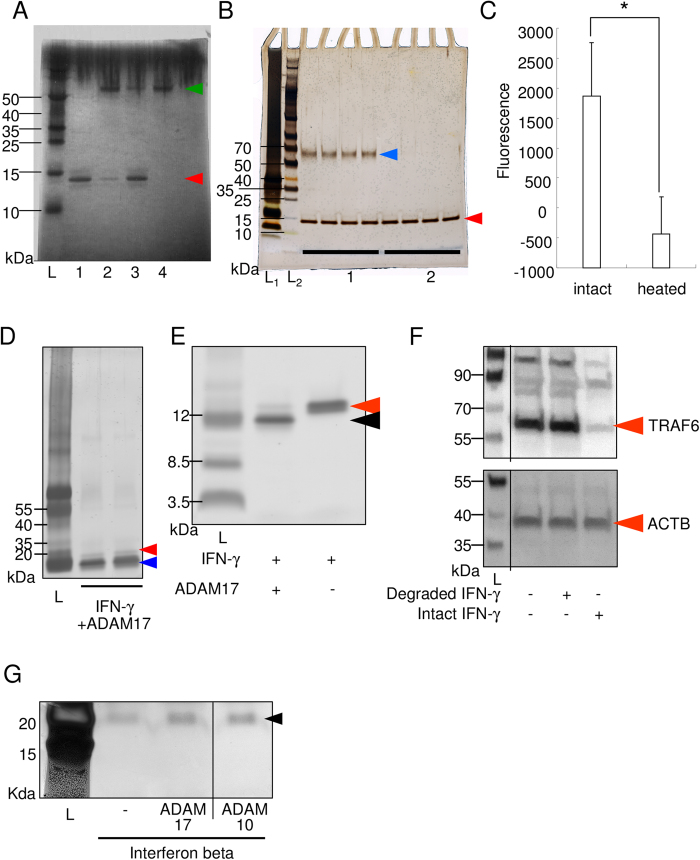
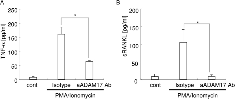
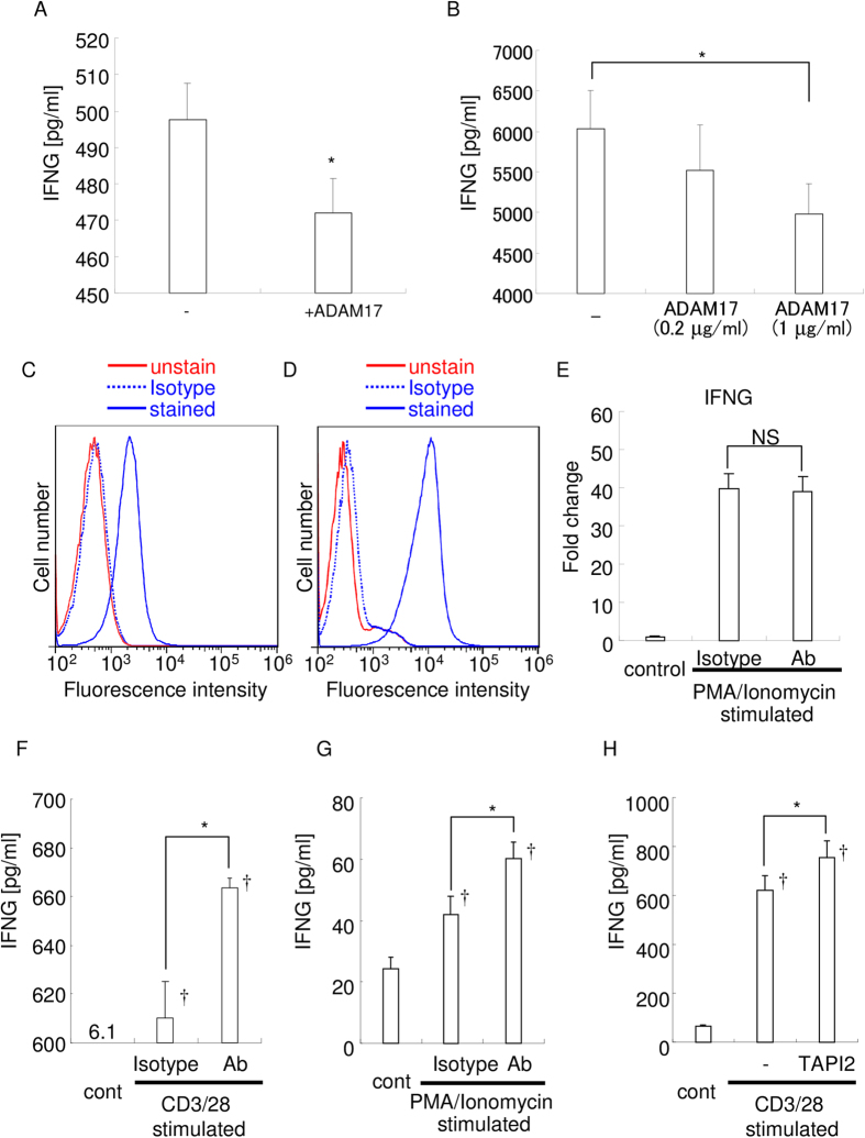
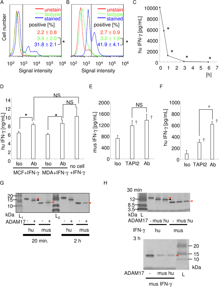
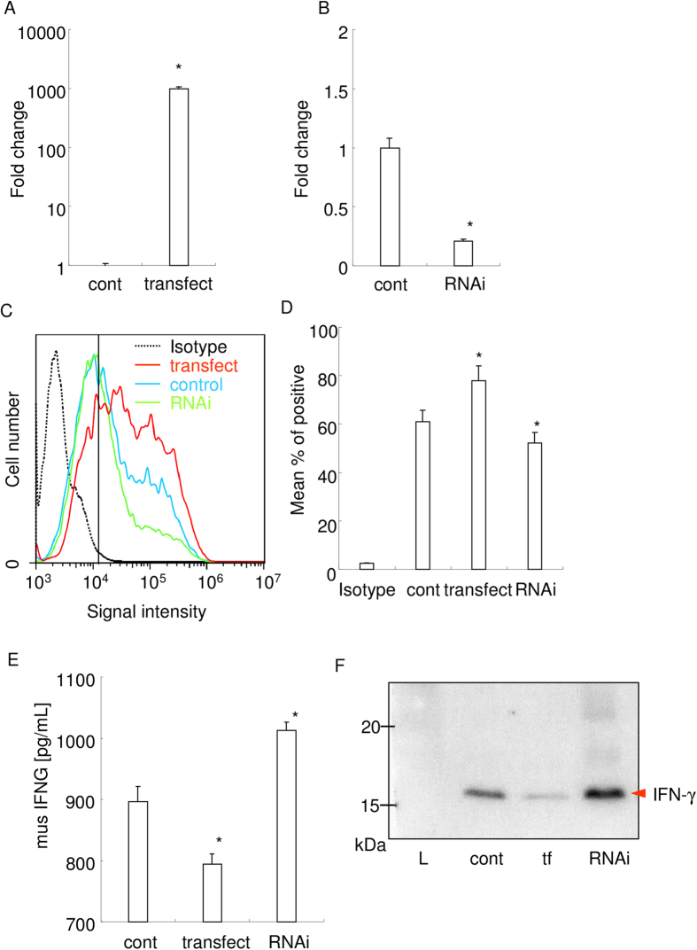
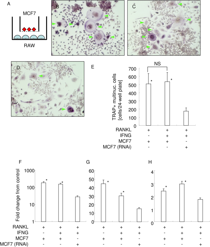
Interferon-gamma (IFN-γ) is a pleiotropic cytokine that exerts anti-tumor and anti-osteoclastogenic effects. Although transcriptional and post-transcriptional regulation of IFN-γ is well understood, subsequent modifications of secreted IFN-γ are not fully elucidated. Previous research indicates that some cancer cells escape immune surveillance and metastasize into bone tissue by inducing osteoclastic bone resorption. Peptidases of the a-disintegrin and metalloproteinase (ADAM) family are implicated in cancer cell proliferation and tumor progression. We hypothesized that the ADAM enzymes expressed by cancer cells degrades IFN-γ and attenuates IFN-γ-mediated anti-tumorigenic and anti-osteoclastogenic effects. Recombinant ADAM17 degraded IFN-γ into small fragments. The addition of ADAM17 to the culture supernatant of stimulated mouse splenocytes decreased IFN-γ concentration. However, ADAM17 inhibition in the stimulated mouse T-cells prevented IFN-γ degradation. ADAM17-expressing human breast cancer cell lines MCF-7 and MDA-MB-453 also degraded recombinant IFN-γ, but this was attenuated by ADAM17 inhibition. Degraded IFN-γ lost the functionality including the inhibititory effect on osteoclastogenesis. This is the first study to demonstrate the extracellular proteolytic degradation of IFN-γ by ADAM17. These results suggest that ADAM17-mediated degradation of IFN-γ may block the anti-tumorigenic and anti-osteoclastogenic effects of IFN-γ. ADAM17 inhibition may be useful for the treatment of attenuated cancer immune surveillance and/or bone metastases.


Figures

Figure 1
Figure 1. ADAM17, but not ADAM10, degrades IFN-γ.
(A) ADAM17 reaction with IFN-γ (Coomassie blue staining of the PAGE gel). The green arrowhead indicates the size of recombinant ADAM17 (rADAM17) (52 kDa), and the red arrowhead indicates the intact recombinant IFN-γ (rIFN-γ) (15.6 kDa). L, molecular weight marker; 1, rIFN-γ; 2, rIFN-γ+rADAM17; 3, rIFN-γ+heat-inactivated rADAM17; 4, rADAM17. A representative photograph is shown. The size of molecular weight marker is indicated on the left side. (B) ADAM10 reaction with IFN-γ (Coomassie blue staining). The blue arrowhead indicates the size of the recombinant ADAM10 (rADAM10: 60kDa), and the red arrowhead indicates recombinant IFN-γ (rIFN-γ). 1, rIFN-γ+rADAM10; 2, rIFN-γ (n = 4 each). (C) Measurement of functional activity of recombinant ADAM10 using fluorescent substrate. Intact or heat inactivated ADAM10 (100 ng) and fluorescent substrate (ES-010; R&D systems: 5 nmol) were incubated at 37 °C and fluorescence was measured at excitation 320 nm and emission 405 nm. *p < 0.05 between the groups. (D) Silver staining of the PAGE gel. The red arrowhead indicates intact recombinant human IFN-γ (hIFN-γ) (15.6 kDa), and blue arrowhead indicates degraded small fragments of hIFN-γ. The fragment indicated by the blue arrowhead was excised and subjected to nano LC-MS/MS analysis. (E) Silver staining of the PAGE gel. IFN-γ with and without ADAM17 were incubated for the same period, and electrophoresed. Red arrowhead indicates intact IFN-γ and the black arrowhead indicates degraded fragment. (F) Functional assay of degraded IFN-γ fragment using western blot analysis for TRAF6 and loading control, ACTB. Separate lanes from the same blot had been spliced together, and lines indicate the places at which the lanes were joined. (G) IFN beta was not degraded by ADAM10 or ADAM17. Recombinant human IFN beta (300 ng) was incubated with or without recombinant ADAM10 or ADAM17 (100 ng) in reaction buffer for 2 hours. Then samples were reduced, denatured, and electrophoresed in TGX precast gel, and silver staining was performed. Arrowhead indicates the size of intact recombinant human IFN beta (22.5 kDa). Separate lanes from the same gel had been spliced together, and lines indicate the places at which the lanes were joined.
Figure 2
Figure 2. Anti-ADAM17 monoclonal antibody.
(A) Inhibition activity of the anti-ADAM17 mAb (100 ng/mL) against TNF-α release. *p < 0.05 between the groups. (B) Inhibition activity of the anti-ADAM17 mAb (100 ng/mL) against sRANKL release. *p < 0.05 between the groups.
Figure 3
Figure 3. Addition of ADAM17 decreases the concentration of IFN-γ and vice versa.
(A) Recombinant IFN-γ (500 pg/ml) was incubated with or without rADAM17 for 2 h, and IFN-γ concentration was measured by ELISA. −, without ADAM17; +ADAM17, with ADAM17; *p < 0.05. (B) Supernatants of PMA/Ionomycin-stimulated mouse splenocytes were incubated with rADAM17 for 2 h, and IFN-γ concentrations were measured by ELISA. *p < 0.05 between the groups. The expression of ADAM17 by CD3/28-stimulated mouse splenocytes (C) and PMA/Ionomycin-stimulated EL4-TK cells (D) was examined by flow cytometry. The red line indicates fluorescence of unstained cells, and the blue line shows fluorescence of cells stained with the CF647-conjugated anti-ADAM17 antibody. Blue dotted line showed the fluorescence of cells stained with the CF647-conjugated isotype IgG. The results are representative of three independent experiments. (E) Real-time PCR analysis of IFN-γ expression. Results are representative of three independent experiments. NS: no statistical difference between the groups. (F) Concentration of the intact IFN-γ in the culture supernatant of CD3/28-stimulated mouse splenocytes. *p < 0.05 between the groups; p < 0.05 versus control. (G) Concentration of intact IFN-γ in the culture supernatant of PMA/Ionomycin-stimulated EL4-TK cells. *p < 0.05 between the groups; p < 0.05 versus control. (H) Concentration of the intact IFN-γ in the culture supernatant of CD3/28-stimulated mouse splenocytes. *p < 0.05 between the groups; p < 0.05 versus control.
Figure 4
Figure 4. ADAM17-expressing breast cancer cells degrade IFN-γ in an ADAM17-dependent manner.
The expression of ADAM17 by MCF7 (A) and MDA-MB-453 (B) cells were examined by flow cytometry. The red line indicates fluorescence of unstained cells, the dotted green line indicates fluorescence of CF647-conjugated isotype IgG stained cells, and the blue line shows fluorescence of stained cells, respectively. The results are representative of three independent experiments, and the mean percentage of ADAM17-positive cells is also shown. The vertical black line indicates the threshold. *p < 0.05 between the groups. (C) Time-course degradation of IFN-γ by MCF7 cells. IFN-γ concentration at each time point was measured. The representative result of three independent experiments is shown. *p < 0.05 versus 0 h. (D–F) ADAM17 blockade inhibited IFN-γ degradation. by MCF7 and MDA-MB-453 cells at 0.5 h. Recombinant human IFN-γ degradation by MCF7 and MDA-MB-453 cells at 0.5 h were shown (D). Recombinant mouse (E) or human (F) IFN-γ degradation by MCF7 cells at 3 h were shown. In the cell culture, isotype control IgG, anti-ADAM17 neutralizing antibody (100 ng/mL), or TAPI2 (10μM) was applied, respectively. *p < 0.05 between the groups. p < 0.05 versus isotype control IgG applied sample. NS: no statistically significant difference between the groups. (G) Comparison of ADAM17-mediated degradation of human and mouse IFN-γ. IFN-γ (200 ng) and ADAM17 (100 ng) was incubated for 20 min or 2 h, electrophoresed, and silver staining was performed. Red arrowhead indicates intact band, and the black arrowhead indicates degraded band. (H) Comparison of IFN-γ degradation activity of human and mouse ADAM17. Human or mouse ADAM17 (100 ng) was incubated with human or mouse IFN-γ (200 ng each) for 30 min or 3 h, electrophoresed, and silver staining was performed. Red arrowhead indicates intact band, and the black arrowhead indicates degraded band.
Figure 5
Figure 5. ADAM17 expressed on MCF-7 dose dependently degrades recombinant IFN-γ.
(A,B) Real-time PCR analysis of ADAM17 mRNA expression in overexpressed or knocked down MCF-7. Fold changes from control were shown. *p < 0.05 versus control. ©Protein level expression of ADAM17 on the cell surface of MCF-7. Fc receptor blocking solution (Biolegend, San Diego, CA) was used prior to antibody application. The results of flow cytometry analysis are shown. Black dotted line indicates the result of fluorophore-conjugated isotype IgG. Blue, red, and green lines indicate the result of control cells, overexpressed cells, and knocked down cells, respectively. (D) Mean percent of positive cells from the results of flowcytometry analysis are shown. *p < 0.05 versus control. (E) IFN-γ concentration measured by ELISA. 1 ng/mL of recombinant mouse IFN-γ were added into each culture media, and incubated for 1 hour. *p < 0.05 versus control. (F) Western blot analysis of IFN-γ performed using the collected culture supernatant. tf: overexpressed cells. RNAi: knocked down cells. L: molecular weight marker.
Figure 6
Figure 6. Biological function analysis of ADAM17-mediated IFN-γ degradation.
(A) Schematic illustration of indirect coculture between RAW 264.7 cells and MCF7 cells. ϕ 1μm cell culture filter insert was used. TRAP staining of RAW 264.7 cells coculture with MCF7 cells in the presence of RANKL (B), coculture with MCF7 cells in the presence of RANKL and IFN-γ (C), and coculture with ADAM17-knockeddown MCF7 cells in the presence of RANKL and IFN-γ (D) are shown, respectively. Green arrowhead indicates TRAP+ multinucleated cells. (E) The number of TRAP+ multinucleated cells in each group are shown. *p < 0.05 vs coculture with ADAM17-knockeddown MCF7 cells in the presence of RANKL and IFN-γ. NS: no significant difference between the groups. Real-time PCR analysis for osteoclast marker gene expression. Fold change from the control of TRAP (F), ATP6v0d2 (G), and cathepsin K (H) were shown, respectively. *p < 0.05 VS coculture with ADAM17-knockeddown MCF7 cells in the presence of RANKL and IFN-γ.

Similar articles

Cited by

References

    1. Dunn G. P., Koebel C. M. & Schreiber R. D. Interferons, immunity and cancer immunoediting. Nature reviews. Immunology 6, 836–848, doi: 10.1038/nri1961 (2006). - DOI - PubMed
    1. Smyth M. J., Hayakawa Y., Takeda K. & Yagita H. New aspects of natural-killer-cell surveillance and therapy of cancer. Nature reviews. Cancer 2, 850–861, doi: 10.1038/nrc928 (2002). - DOI - PubMed
    1. Pestka S., Krause C. D. & Walter M. R. Interferons, interferon-like cytokines, and their receptors. Immunological reviews 202, 8–32, doi: 10.1111/j.0105-2896.2004.00204.x (2004). - DOI - PubMed
    1. Kaplan D. H. et al.. Demonstration of an interferon gamma-dependent tumor surveillance system in immunocompetent mice. Proceedings of the National Academy of Sciences of the United States of America 95, 7556–7561 (1998). - PMC - PubMed
    1. Ishikawa T. et al.. Whole blood interferon-gamma levels predict the therapeutic effects of adoptive T-cell therapy in patients with advanced pancreatic cancer. International journal of cancer. Journal international du cancer, doi: 10.1002/ijc.28117 (2013). - DOI - PubMed

Publication types

MeSH terms

LinkOut - more resources