. 2016 Jul 11;1(8):16108.
doi: 10.1038/nmicrobiol.2016.108.

The binary toxin CDT enhances Clostridium difficile virulence by suppressing protective colonic eosinophilia

Affiliations

The binary toxin CDT enhances Clostridium difficile virulence by suppressing protective colonic eosinophilia

Carrie A Cowardin et al. Nat Microbiol. .

Abstract

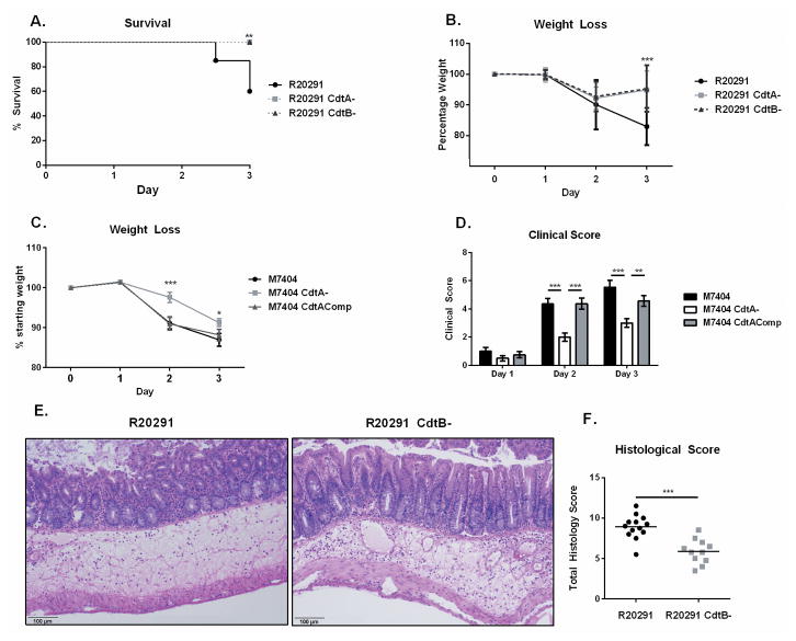
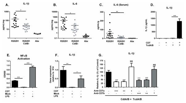
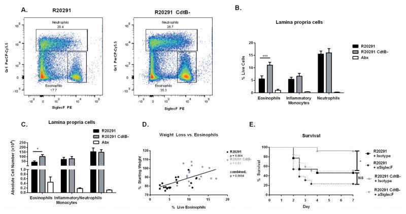
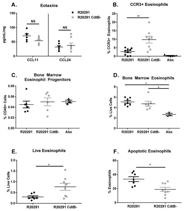
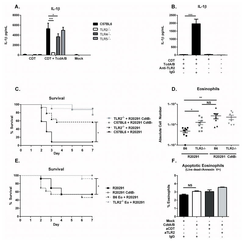
Clostridium difficile is the most common hospital acquired pathogen in the USA, and infection is, in many cases, fatal. Toxins A and B are its major virulence factors, but expression of a third toxin, known as C. difficile transferase (CDT), is increasingly common. An adenosine diphosphate (ADP)-ribosyltransferase that causes actin cytoskeletal disruption, CDT is typically produced by the major, hypervirulent strains and has been associated with more severe disease. Here, we show that CDT enhances the virulence of two PCR-ribotype 027 strains in mice. The toxin induces pathogenic host inflammation via a Toll-like receptor 2 (TLR2)-dependent pathway, resulting in the suppression of a protective host eosinophilic response. Finally, we show that restoration of TLR2-deficient eosinophils is sufficient for protection from a strain producing CDT. These findings offer an explanation for the enhanced virulence of CDT-expressing C. difficile and demonstrate a mechanism by which this binary toxin subverts the host immune response.


Figures

Figure 1
Figure 1. CDT expression enhances the virulence of PCR-ribotype 027 C. difficile in a murine model of infection
(A–B) 8 week old C57BL/6J mice underwent an antibiotic regimen prior to infection with 107 CFU of vegetative C. difficile strain R20291 or the isogenic mutants R20291 CdtA- or R20291 CdtB- (data shown combined from two independent experiments, n=15). (A) Animals were monitored for survival and (B) weight loss. (C–D) Mice were treated with the same antibiotic regimen before infection with 2×10⁵ CFU of M7404, M7404 CdtA- or M7404 CdtAComp (data shown combined from two independent experiments (n=16). (C) Animals were monitored for weight loss and (D) clinical score. (E–F) Mice were sacrificed on day 2 of infection and cecal sections were isolated and fixed in Bouin’s solution for 18 hours before undergoing paraffin embedding, sectioning and haematoxylin & eosin staining. (F) Samples were scored blinded by 3 independent observers. (E) data shown are representative or (F) combined from two independent experiments (n=13). * = p value < 0.05, ** = p value < 0.01, *** = p value < 0.001 by Kaplan-Meier curve (A), two-tailed t-test (B–C) and Mann-Whitney test (D,F). NS = not significant. Error bars shown represent S.D. (B,C) or S.E.M. (D,F).
Figure 2
Figure 2. CDT promotes host inflammatory signaling
(A–B) Mice infected with the indicated strain or uninfected and treated with antibiotics only (Abx) were sacrificed on day 3 of infection and cecal cytokines assessed by lysing whole cecal sections and quantifying protein via ELISA (data combined from two independent experiments, shown normalized to total protein concentration, n=16). (C) Serum IL-6 was measured via ELISA at the same time point (n=14). (D) Bone marrow derived dendritic cells were treated with 200 ng/mL purified CDTa and CDTb (CDT) or 2 ng/mL Toxin A and 2 ng/mL Toxin B (TcdA/B) for 24 hours. Secreted IL-1β was measured by ELISA. (E) NFκB activation was detected in a Raw Blue NFκB reporter cells by measuring Secreted Embryonic Alkaline Phosphatase (SEAP) in the culture media. (F) BMDCs were treated with 200 ng/mL CDTa and 200 ng/mL CDTb or with 100 ng/mL LPS as a positive control for 8 hours. Pro-IL-1β gene expression was assessed by qRT-PCR and is shown normalized to S14 housekeeping gene. (G) BMDCs were treated with 200 ng/mL CDTa and 200 ng/mL CDTb plus 2 ng/mL Toxin A and 2 ng/mL Toxin B in combination with decreasing amounts of anti-CDTa nanobody or anti-CDTb nanobody as indicated. +++ = 200 ng/mL, ++ = 20 ng/mL, + = 2 ng/mL. Secreted IL-1β was measured by ELISA. Data shown combined from 3 independent experiments with 3 replicates each (DG). * = p value < 0.05, ** = p value < 0.01, *** = p value < 0.001 by Welch’s unequal variance t-test (AF) or Mann-Whitney test (G). NS = not significant. Error bars shown represent S.E.M.
Figure 3
Figure 3. CDT production suppresses protective colonic eosinophilia
(A) Mice infected with the indicated strain, or uninfected and treated with antibiotics only (Abx) were sacrificed on day 3. Colon tissue was isolated and processed to a single cell suspension and stained for flow cytometry. Representative flow plots depicting neutrophils and eosinophils are shown (two independent experiments, n=10). (B–C) Eosinophils (CD45+ CD11b+ SiglecF+), monocytes (CD45+ CD11b+ Ly6Chi) and neutrophils (CD45+ CD11b+ Ly6G+) were quantified. All three cell types are significantly elevated in both infected groups compared to Abx treated controls (data shown combined from two independent experiments, n=10). (D) Weight loss and percentage live colon eosinophils were compared using data combined from four independent experiments (n=17). (E) Colonic eosinophils were depleted using 40 ug of an anti-SiglecF targeted antibody or an isotype control antibody one day prior and one day following infection with C. difficile. Animals were monitored for clinical symptoms and humanely euthanized when required. Data shown combined from two independent experiments (n = 13). * = p value < 0.05, ** = p value < 0.01, *** = p value < 0.001 by Mann-Whitney test (B–C), linear regression (D) or Kaplan-Meier curve (E). NS = not significant. Error bars shown represent S.E.M.
Figure 4
Figure 4. CDT production by C. difficile promotes eosinophil apoptosis
(A) Mice infected with the indicated strain, or uninfected and treated with antibiotics (Abx) were sacrificed on day 3. Cecal eotaxin levels were assessed by lysing whole cecal sections and quantifying protein via ELISA (data combined from two independent experiments, shown normalized to total protein concentration, n=16). (B) Colon tissue was isolated and processed to a single cell suspension and stained for flow cytometry. CCR3 staining on eosinophils was assessed by first gating on total eosinophils (identified as CD45+ CD11b+ SiglecF+ SSChi cells) (data shown are representative of 2 independent experiments, n=8). (C–D) Mice were sacrificed on day 3 and bone marrow harvested for flow cytometry. Eosinophil progenitors were identified as Lin- CD34+ Sca-1 IL-5Rα+ cKitint cells by flow cytometry. Mature bone marrow eosinophils were quantified as CD45+ CD11b+ SiglecF+ SSChi cells (data shown combined from 2 independent experiments, n=6). (E–F) Blood eosinophils were assessed on day 3 of infection by flow cytometry following cardiac puncture, red blood cell lysis, and staining. Live eosinophils were identified at CD45+ CD11b+ SiglecF+ Live deadneg and apoptotic eosinophils identified as CD45+ CD11b+ SiglecF+ Annexin V+ Live deadneg cells. Data shown combined from two independent experiments. (n=7) * = p value < 0.05, ** = p value < 0.01, *** = p value < 0.001 by Mann-Whitney test (A, C–D, F) or Welch’s unequal variance t-test (B, E). NS = not significant. Error bars shown represent S.E.M.
Figure 5
Figure 5. TLR2 mediates CDT recognition and is required for eosinophil suppression
(A) BMDCs were generated from TLR2−/−, TLR4−/−, and TLR5−/− mice before treatment with 200 ng/mL CDTa and 200 ng/mL CDTb (CDT) and 2 ng/mL Toxin A and 2 ng/mL Toxin B (TcdA/B) for 24 hours. IL-1β secretion was assessed by ELISA. (B) BMDCs were treated with 200 ng/mL CDTa and 200 ng/mL CDTb (CDT) and 2 ng/mL Toxin A and 2 ng/mL Toxin B (TcdA/B) for 24 hours in the presence of a TLR2 neutralizing antibody or an isotype control. IL-1β was assessed by ELISA. Data shown combined from three independent experiments of 3 replicates each (AB). (C) 8 week old TLR2 knockout mice or C57BL/6J mice were infected with R20291 or R20291 CdtB- and monitored for survival (data combined from two independent experiments, n=14). (D) Mice were sacrificed on day 3 of infection and colonic eosinophils (CD45+ CD11b+ SiglecF+ SSChi) were measured by flow cytometry following tissue processing and staining (data combined from two independent experiments, n=8). (E) C57BL/6 mice received 4×10⁵ TLR2−/− or B6 bone marrow-derived eosinophils (TLR2−/− Eo or B6 Eo) via IP injection one day prior and for 3 subsequent days following infection with R20291 or R20291 CdtB-. Mice were monitored daily for survival (data combined from two independent experiments, n=13). (F) BM Eos were incubated for 8 hours with 200 ng/mL CDTa and 200 ng/mL CDTb in the presence or absence of anti-TLR2 neutralizing antibody (aTLR2) or anti-CDT neutralizing nanobody (aCDT). Eosinophils were stained with Live dead or Annexin V and cell death was assessed by flow cytometry, data shown are representative of 3 independent experiments assayed in duplicate. * = p value < 0.05, ** = p value < 0.01, *** = p value < 0.001 by Mann-Whitney test (A), Welch’s unequal variance t-test (B, D, F) or Kaplan-Meier Analysis (C, E). NS = not significant. Error bars shown represent S.E.M.

Similar articles

Cited by

References

    1. Lessa FC, et al. Burden of Clostridium difficile infection in the United States. N Engl J Med. 2015;372:825–834. - PMC - PubMed
    1. Ananthakrishnan AN. Clostridium difficile infection: epidemiology, risk factors and management. Nat Rev Gastroenterol Hepatol. 2010;8:17–26. - PubMed
    1. Kuehne SA, et al. The role of toxin A and toxin B in Clostridium difficile infection. Nature. 2010;467:711–713. - PubMed
    1. Carter GP, Rood JI, Lyras D. The role of toxin A and toxin B in Clostridium difficile-associated disease. Gut Microbes. 2010;1:58–64. - PMC - PubMed
    1. Lyerly DM, Saum KE, MacDonald DK, Wilkins TD. Effects of Clostridium difficile toxins given intragastrically to animals. Infect Immun. 1985;47:349–352. - PMC - PubMed

MeSH terms

Substances

LinkOut - more resources