. 2016 Aug 30:6:32203.
doi: 10.1038/srep32203.

Decreased demand for olfactory periglomerular cells impacts on neural precursor cell viability in the rostral migratory stream

Affiliations

Decreased demand for olfactory periglomerular cells impacts on neural precursor cell viability in the rostral migratory stream

Anika Langenfurth et al. Sci Rep. .

Abstract

The subventricular zone (SVZ) provides a constant supply of new neurons to the olfactory bulb (OB). Different studies have investigated the role of olfactory sensory input to neural precursor cell (NPC) turnover in the SVZ but it was not addressed if a reduced demand specifically for periglomerular neurons impacts on NPC-traits in the rostral migratory stream (RMS). We here report that membrane type-1 matrix metalloproteinase (MT1-MMP) deficient mice have reduced complexity of the nasal turbinates, decreased sensory innervation of the OB, reduced numbers of olfactory glomeruli and reduced OB-size without alterations in SVZ neurogenesis. Large parts of the RMS were fully preserved in MT1-MMP-deficient mice, but we detected an increase in cell death-levels and a decrease in SVZ-derived neuroblasts in the distal RMS, as compared to controls. BrdU-tracking experiments showed that homing of NPCs specifically to the glomerular layer was reduced in MT1-MMP-deficient mice in contrast to controls while numbers of tracked cells remained equal in other OB-layers throughout all experimental groups. Altogether, our data show the demand for olfactory interneurons in the glomerular layer modulates cell turnover in the RMS, but has no impact on subventricular neurogenesis.


Figures

Figure 1
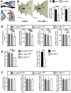
Figure 1. MT1-MMP deficiency affects the structure of olfactory turbinates and the level of olfactory bulb sensory innervation.
(A) Overview on the olfactory epithelium (OE, foldings are termed turbinates) and olfactory bulb (OB); enlarged part shows olfactory sensory neurons and glomeruli; white area between olfactory epithelium and olfactory bulb is the cribriform plate; olfactory sensory neurons are abbreviated as OSN; the black and white arrow indicates olfactory glomeruli. (B,C) Turbinates in MT1-MMP−/− were less complex than in controls, the olfactory epithelium (OE) was labelled with the olfactory marker protein and is visible in dark brown colour. (D) The surface area of the nasal turbinates is strongly reduced in MT1-MMP−/− mice (white bar) as compared to wildtype controls (black bar; p = 0.0108) (E,F) Nasal cavity and anterior olfactory bulb of olfactory receptor mOR256-17 reporter mice (mOR256-17-GFP crossed with MT1-MMP−/− or control): olfactory sensory neurons (arrowhead) pass through the cribriform plate (asterisk) and into a defined glomerulus (inserts in D,E); Nuclei are labeled with DAPI in blue. (G) The number of mOR256-17-GFP+ neurons was reduced in MT1-MMP−/− mice. Scale bars: 200 μm in (B,C). 100 μm in (E,F); 100 μm in inserts (E,F); statistical significance: *represents p < 0.05; **represents p < 0.005.
Figure 2
Figure 2. MT1-MMP deficiency affects the size of the olfactory bulb.
(A) A schematic drawing shows the position of the frontal forebrain and olfactory bulb (OB) in relation to the nasal cavity (upper part); lower part: a horizontal section through the OB (blue structure) is magnified and the position of the olfactory glomeruli (red circles) is highlighted (arrowhead). (B) Phase contrast images of a sagittal section through the OB in MT1-MMP−/− and controls. (C) Olfactory bulb size is largely reduced in MT1-MMP−/− at P20; a reduction in OB-size is not associated with increased numbers for myeloid cells (Iba1+ cells) in the OB. (D) Histochemical staining and subsequent cell-counting were used to quantify the total number of cells per olfactory glomerulus in conditional MT1-MMP knockouts (MT1-MMPΔ/Δ) or controls (MT1-MMPΔ/WT, Mt1-MMPFL/FL); immunohistochemical staining for calretinin, tyrosine-hydroxylase (TH) or calbindin was followed by cell-counting to quantify numbers of interneuron subtypes in the glomerular cell layer. (E) Histochemical staining and morphometry were used to quantify the size of glomeruli in all mouse models. (F) Histochemistry or immunohistochemistry and cell-counting were performed to quantify the number of cells respectively the number of different interneuron in the glomerular cell layer. Scale bars: 1 mm in (B,C); statistical significance: ** represents p < 0.005; *** represents p < 0.001.
Figure 3
Figure 3. MT1-MMP deficiency has no effect on subventricular neurogenesis.
(A) Scheme indicating the anatomical position of the subventricular zone (SVZ) in a sagittal brain section. (B,C) Immunohistochemistry for BrdU (labelled cells are magnified and shown in the insert; a 2 h BrdU-pulse was applied) in the SVZ; labelling for DNA strand breaks (TUNEL) indicating cell-death in the SVZ; representative staining for proliferative and dying subventricular cells in MT1-MMP−/− mice and controls are shown. (C) Quantification of total cell numbers for BrdU+, Ki67+ and TUNEL+ cells in the SVZ of MT1-MMP knockouts and controls. Scale bars: 100 μm (BrdU) and 5 μm (for insert in BrdU), 10 μm (TUNEL); statistical significance: ***represents p < 0.001; *represents p < 0.05.
Figure 4
Figure 4. Reduced numbers of new periglomerular cells and increased cell-death levels in the RMS of MT1-MMP deficient mice.
(A) Scheme indicating the localization of the RMS in a sagittal brain section and in a magnified horizontal section of the migratory path in the OB. (B) Immunohistochemistry for BrdU was performed on a horizontal OB-section: granule cell-layer (GR), external plexiform layer (EPL), glomerular layer (GL). (C,D) Representative staining for BrdU. (E) The number of BrdU+ cells (labelling protocol as described in B) was quantified in distinct layers of the olfactory bulb using horizontal sections of identical stereotactic coordinates in MT1-MMPΔ/Δ or controls (summarized from MT1-MMPΔ/WT and MT1-MMPFL/FL experimental groups, which gave highly similar results). (F) Composite pictures showing representative immunolabelling for the proliferation marker Ki67 in the subventricular zone (SVZ) and rostral extension of the SVZ plus rostral migratory stream (divided into the parts 1, 2 and 3) of MT1-MMP−/− and control mice. (G) Quantification of Ki67+ and TUNEL+ cells throughout the anatomical areas highlighted in F as 1, 2 and 3; note that cell-death levels in the RMS (area 2 or 3) are robustly increased in MT1-MMP deficient mice as compared to controls while proliferation-rates remain unchanged. (H) MT1-MMPΔ/Δ mice or controls received repetitive BrdU-pulse followed by an extended chase period (for details see text) and sagittal brain sections were subsequently immunolabelled for DCX (green) and BrdU (red). Overviews on parts 2 and 3 of the RMS (indicated by arrowheads) are provided. Cells in the RMS were inspected by confocal microscopy and analysed for coexpression of markers (crosshair images); single optical sections and corresponding immunofluorescence over 4 μm in Z-orientation are presented; note that BrdU+/DCX+ cells can be clearly identified. (I) The number of DCX and BrdU colabelled cells was quantified in the proximal (area-1), intermediate (area-2) and distal (area-3) part of the RMS in MT1-MMP deficient and control mice. Scale bars: 250 μm in (B), 25 μm in (C,D) 1 mm in (F) and 500 μm in (H); statistical significance: n.s. indicates no statistical significance; *represents p < 0.05; ***represents p < 0.001.

References

    1. Kriegstein A. & Alvarez-Buylla A. The glial nature of embryonic and adult neural stem cells. Annu Rev Neurosci 32, 149–184, doi: 10.1146/annurev.neuro.051508.135600 (2009). - DOI - PMC - PubMed
    1. Kronenberg G. et al.. Impact of actin filament stabilization on adult hippocampal and olfactory bulb neurogenesis. The Journal of neuroscience: the official journal of the Society for Neuroscience 30, 3419–3431, doi: 10.1523/JNEUROSCI.4231-09.2010 (2010). - DOI - PMC - PubMed
    1. Alvarez-Buylla A. & Lim D. A. For the long run: maintaining germinal niches in the adult brain. Neuron 41, 683–686 (2004). - PubMed
    1. Lledo P. M., Alonso M. & Grubb M. S. Adult neurogenesis and functional plasticity in neuronal circuits. Nat Rev Neurosci 7, 179–193 (2006). - PubMed
    1. Brill M. S. et al.. Adult generation of glutamatergic olfactory bulb interneurons. Nature neuroscience 12, 1524–1533, doi: 10.1038/nn.2416 (2009). - DOI - PMC - PubMed

Publication types

MeSH terms

Substances

LinkOut - more resources