. 2016 Sep 13;113(37):10316-21.
doi: 10.1073/pnas.1607210113. Epub 2016 Aug 29.

Anatomy of the β-branching enzyme of polyketide biosynthesis and its interaction with an acyl-ACP substrate

Affiliations

Anatomy of the β-branching enzyme of polyketide biosynthesis and its interaction with an acyl-ACP substrate

Finn P Maloney et al. Proc Natl Acad Sci U S A. .

Abstract

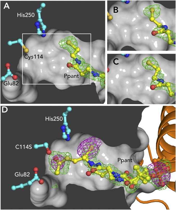
Alkyl branching at the β position of a polyketide intermediate is an important variation on canonical polyketide natural product biosynthesis. The branching enzyme, 3-hydroxy-3-methylglutaryl synthase (HMGS), catalyzes the aldol addition of an acyl donor to a β-keto-polyketide intermediate acceptor. HMGS is highly selective for two specialized acyl carrier proteins (ACPs) that deliver the donor and acceptor substrates. The HMGS from the curacin A biosynthetic pathway (CurD) was examined to establish the basis for ACP selectivity. The donor ACP (CurB) had high affinity for the enzyme (Kd = 0.5 μM) and could not be substituted by the acceptor ACP. High-resolution crystal structures of HMGS alone and in complex with its donor ACP reveal a tight interaction that depends on exquisite surface shape and charge complementarity between the proteins. Selectivity is explained by HMGS binding to an unusual surface cleft on the donor ACP, in a manner that would exclude the acceptor ACP. Within the active site, HMGS discriminates between pre- and postreaction states of the donor ACP. The free phosphopantetheine (Ppant) cofactor of ACP occupies a conserved pocket that excludes the acetyl-Ppant substrate. In comparison with HMG-CoA (CoA) synthase, the homologous enzyme from primary metabolism, HMGS has several differences at the active site entrance, including a flexible-loop insertion, which may account for the specificity of one enzyme for substrates delivered by ACP and the other by CoA.

Keywords: HMG synthase; acyl carrier protein; curacin; natural products; polyketide synthase.


Conflict of interest statement

The authors declare no conflict of interest.

Figures

Fig. 1.
Fig. 1.
HMGS reaction. (A) Reaction steps. (1) ACPD transfers an acetyl group to HMGS Cys114 and Glu82 deprotonates the acetyl group; (2) the resulting enolate nucleophile attacks acetoacetyl-ACPA; (3) the HMG-ACPA product is hydrolyzed from Cys114. R indicates the polyketide intermediate (methyl in curacin A biosynthesis). (B) Structure of the Ppant cofactor (represented as a squiggle symbol in A).
Fig. S1.
Fig. S1.
Curacin β-branching. (A) CurC KSDC is proposed to decarboxylate malonyl-CurB, generating acetyl-CurB (ACPD), (1) CurD HMGS catalyzes formation of HMG-CurA ACP3 (ACPA), (2) CurA Hal chlorinates the γ-carbon of HMG-ACPA, which is subsequently dehydrated to 3 and decarboxylated to 4 by CurE ECH1 and CurF ECH2, respectively. CurF ER catalyzes cyclopropanation to 6 by NADPH-dependent addition of hydride and elimination of chloride. (B) Reaction catalyzed by HMGCS, the HMGS homolog from primary metabolism.
Fig. S2.
Fig. S2.
Sample HMGS activity data. Ppant ejection mass spectra of the ACPA elution of an HMGS reaction mix. Ion counts were recorded for the acetoacetyl (calculated m/z = 345.15) and HMG (calculated m/z = 405.17) peaks. See Table S3 for total ion counts for each species.
Fig. 2.
Fig. 2.
CurD HMGS structure. Within the dimer, the right-hand subunit is colored by sequence from the blue N terminus to the red C terminus. Key residues are shown in ball-and-stick on the gray left monomer, including the catalytic Cys114, Glu82, and His250. Phe148 and Ala164 are the boundaries of the 15-residue disordered loop at the active site entrance. The basic side chain of Arg33 interacts with the Ppant phosphate and is conserved in HMGCS sequences. A dotted line denotes the disordered loop region connecting Phe148 to Ala164.
Fig. S3.
Fig. S3.
Multiple sequence alignments. Alignments were generated using Clustal Omega (54) and analyzed in Jalview (55). (A) Alignment of HMGS and HMGCS sequences. The boxed sequences on top, identified by protein name, are for the β-branching HMGS and the bottom sequences are for the primary metabolism HMGCS, identified by species of origin. Important residues are numbered by the CurD HMGS sequence. Ppant interacting residues are identified in yellow, catalytic residues in blue, thiol pocket residues in green, and ACPD-interacting residues in red. Species of origin and accession codes for HMGS sequences are as follows: CurD from Moorea producens, Q6DNE9; PksG from Bacillus subtilis, P40830; MupH from Pseudomonas fluorescens, Q8RL63; TaC from Myxococcus xanthus, Q1D5E5; TaF from Myxococcus xanthus, Q1D5E8; BryR from Ca. Endobugula sertula, A2CLL9. Accession codes for HMGCS sequences: Enterococcus faecalis, Q9FD71; Staphylococcus aureus, Q9FD87; Brassica juncea, Q9M6U3; Homo sapiens, Q01581. (B) Alignment of ACPD and ACPA sequences. The alignment was generated using Clustal Omega (54) and analyzed in Jalview (55). Boxed sequences are for ACPD and below for ACPA. Important residues are numbered by the CurB ACPD sequence. The positions of hydrophobic side chains that are conserved aliphatics in ACPD and aromatic in ACPA are indicated by arrows below the alignment (Fig. S6). Species of origin and accession codes for HMGS sequences are as follows: CurB and CurA from Moorea producens 3L (F4Y434, F4Y435); MacpC and MmpA3_ACPs from Pseudomonas fluorescens (Q8RL65, Q8RL76); AcpK and PksL from Bacillus subtilis (Q7PC63, Q05470); TaB, TaE, and Ta1 ACPs from Myxococcus xanthus (Q9XB07, Q9XB04, Q9Z5F4), and BryA and BryC from Candidatus Endobugula sertula (A2CLL5, A2CLL2).
Fig. S4.
Fig. S4.
Electron density for holo-ACPD-HMGS. Key residues (ball-and-stick) were omitted, and models were refined in phenix.refine (56) with simulated annealing from a start temperature of 5,000 °C. SA-omit density is contoured at 3.0σ in green (A–H) and at 1.5σ in pale green (D and F). HMGS residues are in cyan, ACPD in orange, and Ppant in yellow in each panel. (A) Catalytic HMGS residues. (B) Ppant. (C–G) Ionic contacts (Fig. 3C). (H) Nonpolar contacts (Fig 3D). Weaker omit density for Arg33 and Arg266 is shown in D and F.
Fig. 3.
Fig. 3.
HMGS interaction with the donor ACP. (A) ACPD (orange)/HMGS (cyan) complex. Ppant (yellow) and catalytic residues shown are in ball-and-stick form. (B) Ppant in the HMGS active site. Dashed yellow lines represent hydrogen bonds and the long separation of Ppant and Cys114 thiol groups. (C) Stereoview of charged contacts in the HMGS-ACPD interface. (D) Stereoview of hydrophobic contacts between ACPD and HMGS. HMGS helices are numbered as in Fig. 2, and ACPD helices are labeled by Roman numerals. Helices in C and D are transparent for clarity.
Fig. 4.
Fig. 4.
Acetylation-dependent position of Ppant. Panels show Fo-Fc omit density (3σ, Ser-Ppant omitted, green) for structures of ACPD-HMGS in different biochemical states crystallized in identical conditions. (A) Holo-ACPD and HMGSWT. White box indicates field of view for B and C. (B) Acetyl-ACPD and HMGSWT, showing that the acetyl group has been lost. (C) Holo-ACPD and HMGSC114S. (D) Acetyl-ACPD and HMGSC114S. Anomalous difference density (3σ, magenta) indicates that S is present in both terminal densities for the Ppant and also shows the Ppant P atom. Atoms are colored as in Fig. 3.
Fig. S5.
Fig. S5.
Affinity of ACPD for WT and variant HMGS. Fluorescence anisotropy of BODIPY-tagged ACPD was recorded as a function of HMGS concentration. For each HMGS variant, binding curves are shown for apo-ACPD (Left) and holo-ACPD (Right). Data were recorded and analyzed with Graphpad Prism. Data represent the average of three measurements.
Fig. S6.
Fig. S6.
Electrostatic surface potentials and interacting surfaces for HMGS and selected ACPs. For ACPs from structures of enzyme complexes, the black outline delineates the molecular surface within 5 Å of any atom in the interacting enzyme and the yellow star denotes the site of Ppant attachment. (A) CurD HMGS (complex with CurB ACPD). (B) CurB holo-ACPD (complex with CurD HMGS). (C) CurA ACPA [2LIW (24); RMSD 2.2 Å]. (D) DEBS module 2 ACP [2JU2 (25); RMSD 2.7 Å]. (E) VinL ACP [5CZD (31); complex with VinK Acyltransferase, RMSD 2.6 Å]. (F) E. coli AcpP [complex with E. coli FabA dehydratase, 4KEH (26); RMSD 2.1 Å]. (G) E. coli AcpP [complex with E. coli LpxD, 4IHG (27); RMSD 3.5 Å]. (H) B. subtilis ACP [complex with B. subtilis ACP synthase, 1F80 (29); RMSD = 2.2 Å]. (I) R. communis ACP [complex with R. communis ACP desaturase, 2XZ1 (28); RMSD 1.7]. Molecular surfaces are colored by electrostatic potential (±5 kT/e, blue electropositive, red electronegative) (57). RMSD values are from Cα superposition with CurB ACPD.

Similar articles

Cited by

References

    1. Cragg GM, Newman DJ. Natural products: A continuing source of novel drug leads. Biochim Biophys Acta. 2013;1830(6):3670–3695. - PMC - PubMed
    1. Weissman KJ. Genetic engineering of modular PKSs: From combinatorial biosynthesis to synthetic biology. Nat Prod Rep. 2016;33(2):203–230. - PubMed
    1. Calderone CT. Isoprenoid-like alkylations in polyketide biosynthesis. Nat Prod Rep. 2008;25(5):845–853. - PubMed
    1. Blokhin AV, et al. Characterization of the interaction of the marine cyanobacterial natural product curacin A with the colchicine site of tubulin and initial structure-activity studies with analogues. Mol Pharmacol. 1995;48(3):523–531. - PubMed
    1. Verdier-Pinard P, et al. Structure-activity analysis of the interaction of curacin A, the potent colchicine site antimitotic agent, with tubulin and effects of analogs on the growth of MCF-7 breast cancer cells. Mol Pharmacol. 1998;53(1):62–76. - PubMed

Publication types

MeSH terms

LinkOut - more resources