

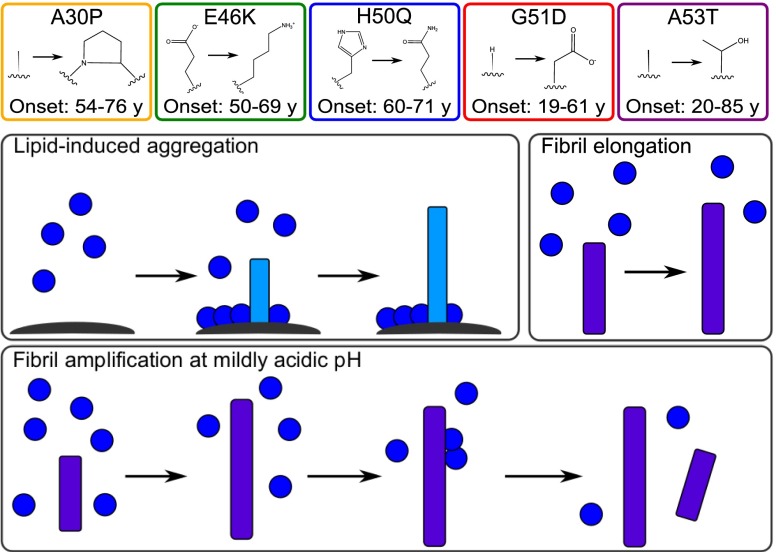
Mutations associated with familial Parkinson's disease alter the initiation and amplification steps of α-synuclein aggregation
- PMID: 27573854
- PMCID: PMC5027465
- DOI: 10.1073/pnas.1604645113
Mutations associated with familial Parkinson's disease alter the initiation and amplification steps of α-synuclein aggregation
Abstract
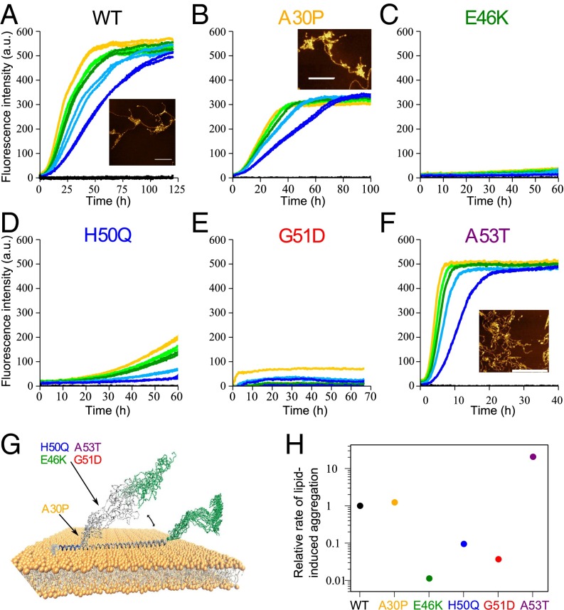
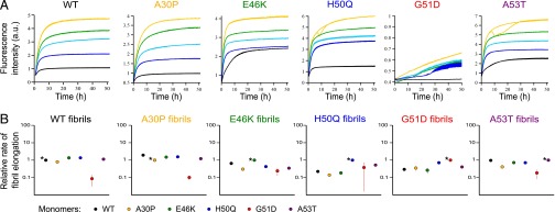
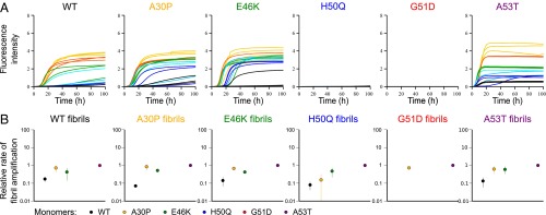
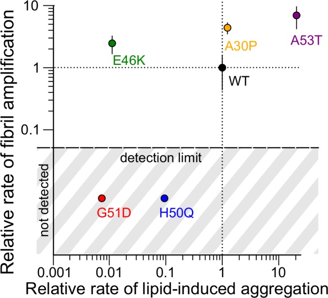
Parkinson's disease is a highly debilitating neurodegenerative condition whose pathological hallmark is the presence in nerve cells of proteinacious deposits, known as Lewy bodies, composed primarily of amyloid fibrils of α-synuclein. Several missense mutations in the gene encoding α-synuclein have been associated with familial variants of Parkinson's disease and have been shown to affect the kinetics of the aggregation of the protein. Using a combination of experimental and theoretical approaches, we present a systematic in vitro study of the influence of disease-associated single-point mutations on the individual processes involved in α-synuclein aggregation into amyloid fibrils. We find that lipid-induced fibril production and surface catalyzed fibril amplification are the processes most strongly affected by these mutations and show that familial mutations can induce dramatic changes in the crucial processes thought to be associated with the initiation and spreading of the aggregation of α-synuclein.
Keywords: familial Parkinson’s disease; kinetic analysis; lipid-induced aggregation; neurodegenerative disease; seeded aggregation.
Conflict of interest statement
The authors declare no conflict of interest.
Figures
References
-
- Nakajo S, et al. Purification and characterization of a novel brain-specific 14-kDa protein. J Neurochem. 1990;55(6):2031–2038. - PubMed
-
- Shibayama-Imazu T, et al. Cell and tissue distribution and developmental change of neuron specific 14 kDa protein (phosphoneuroprotein 14) Brain Res. 1993;622(1-2):17–25. - PubMed
-
- Iwai A, et al. The precursor protein of non-A β component of Alzheimer’s disease amyloid is a presynaptic protein of the central nervous system. Neuron. 1995;14(2):467–475. - PubMed
Publication types
MeSH terms
Substances
Grants and funding
LinkOut - more resources
Full Text Sources
Other Literature Sources
Medical
