Review
. 2016 Aug;6(4):358-368.
doi: 10.1212/CPJ.0000000000000265.

Ultrasound guidance for lumbar puncture

Affiliations
Review

Ultrasound guidance for lumbar puncture

Nilam J Soni et al. Neurol Clin Pract. 2016 Aug.

Abstract

Purpose of review: To review the literature and describe techniques to use ultrasound to guide performance of lumbar puncture (LP).

Recent findings: Ultrasound evaluation of the lumbar spine has been shown in randomized trials to improve LP success rates while reducing the number of attempts and the number of traumatic taps.

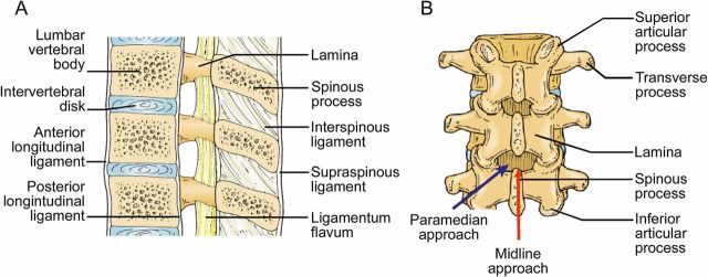
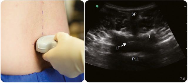
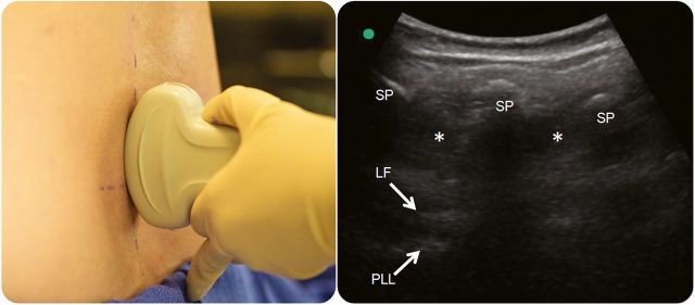
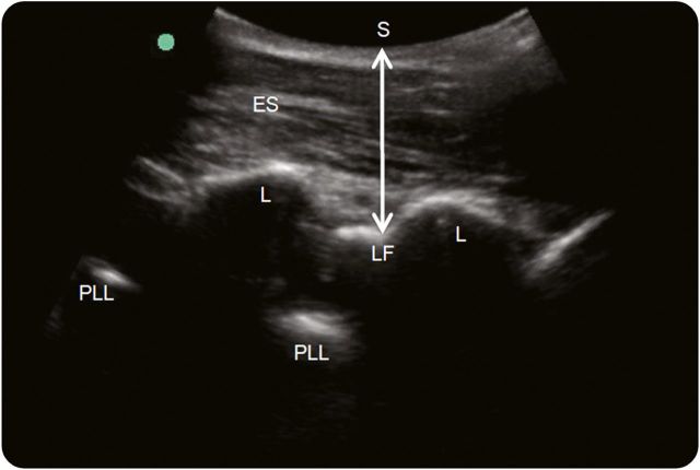
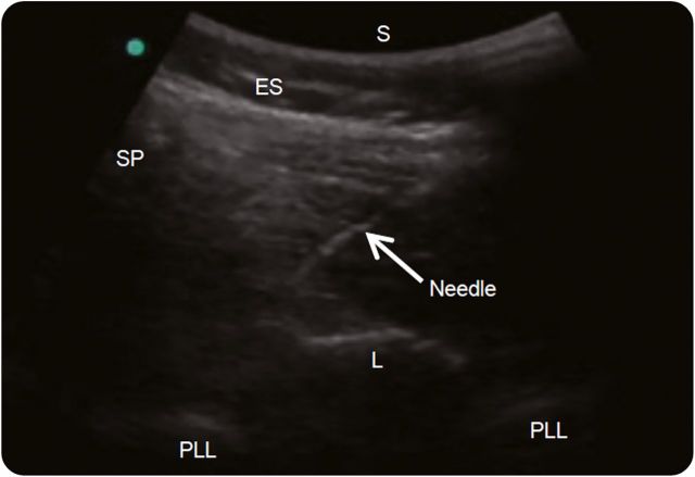
Summary: Ultrasound mapping of the lumbar spine reveals anatomical information that is not obtainable by physical examination, including depth of the ligamentum flavum, width of the interspinous spaces, and spinal bone abnormalities, including scoliosis. Using static ultrasound, the lumbar spine anatomy is visualized in transverse and longitudinal planes and the needle insertion site is marked. Using real-time ultrasound guidance, the needle tip is tracked in a paramedian plane as it traverses toward the ligamentum flavum. Future research should focus on efficient methods to train providers, cost-effectiveness of ultrasound-guided LP, and the role of new needle-tracking technologies to facilitate the procedure.


Figures

Figure 1
Figure 1. Lumbar spine anatomy
(A) Ligaments of the lumbar spine: The supraspinous ligament connects the tips of the spinous processes, the interspinous ligament connects the shafts of the spinous processes, and the ligamentum flavum connects the lamina. The posterior longitudinal ligament is a dense band that stretches along the posterior aspect of the vertebral bodies. (B) Midline vs paramedian approach to lumbar puncture: Using a traditional midline approach, the spinal needle is inserted in the narrow space in between spinous processes, while in a paramedian approach, the spinal needle is inserted lateral to the spinous processes and angled toward the center of the spinal canal. (Reprinted with permission from Point-of-Care Ultrasound by Nilam J. Soni, Robert Arntfield, and Pierre Kory, ch. 35, pp. 283–284, Elsevier Health Sciences, 2014).
Figure 2
Figure 2. Transverse midline view
A lumbar spinous process is centered on the screen with the transducer in a transverse plane, and a mark is made perpendicular to the transducer. Sliding the transducer along the midline allows visualization of the spinous processes (SP), lamina (L), posterior longitudinal ligament (PLL), and ligamentum flavum (LF).
Figure 3
Figure 3. Longitudinal midline view
The transducer is centered over a lumbar interspinous space in a longitudinal plane, and a mark is made perpendicular to the center of the transducer. The spinous processes (SP) and interspinous spaces (*) are visualized in a longitudinal plane, and the ligamentum flavum (LF) and posterior longitudinal ligament (PLL) are visualized deep to the spinous processes.
Figure 4
Figure 4. Paramedian view
With the transducer oriented longitudinally on one side of the midline, the erector spinae (ES) muscles are seen superficial to the lamina (L). From a paramedian view, the ligamentum flavum (LF) is easily visualized, and the skin (S)–LF distance (double-headed arrow) can be measured. The posterior longitudinal ligament (PLL) is seen deep to the ligamentum flavum.
Figure 5
Figure 5. Real-time ultrasound guidance
From an oblique paramedian view of lumbar spine, a spinal needle is inserted using an in-plane technique toward the lamina-ligamentum flavum junction. The transducer is oriented obliquely from the spinous process (SP) of the superior vertebra to the lamina (L) of the inferior vertebra. The needle is inserted underneath the transducer in a lateral to medial direction. ES = erector spinae muscles; PLL = posterior longitudinal ligament; S = skin.

References

    1. Edwards C, Leira EC, Gonzalez-Alegre P. Residency training: a failed lumbar puncture is more about obesity than lack of ability. Neurology 2015;84:e69–e72. - PubMed
    1. Bogin IN, Stulin ID. Application of the method of 2-dimensional echospondylography for determining landmarks in lumbar punctures [in Russian]. Zh Nevropatol Psikhiatr Ime S S Korsakova 1971;71:1810–1811. - PubMed
    1. Grau T, Leipold R, Conradi R, Martin E, Motsch J. Ultrasonography and peridural anesthesia: technical possibilities and limitations of ultrasonic examination of the epidural space [in German]. Anaesthesist 2001;50:94–101. - PubMed
    1. Grau T, Leipold RW, Conradi R, Martin E. Ultrasound control for presumed difficult epidural puncture. Acta Anaesthesiol Scand 2001;45:766–771. - PubMed
    1. Grau T, Leipold RW, Horter J, Conradi R, Martin E, Motsch J. The lumbar epidural space in pregnancy: visualization by ultrasonography. Br J Anaesth 2001;86:798–804. - PubMed