. 2016 Aug 30;5(8):e357.
doi: 10.1038/mtna.2016.72.

(18)F-FDG PET/CT for Monitoring the Response of Breast Cancer to miR-143-Based Therapeutics by Targeting Tumor Glycolysis

Affiliations

(18)F-FDG PET/CT for Monitoring the Response of Breast Cancer to miR-143-Based Therapeutics by Targeting Tumor Glycolysis

Ying Miao et al. Mol Ther Nucleic Acids. .

Abstract

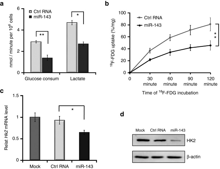
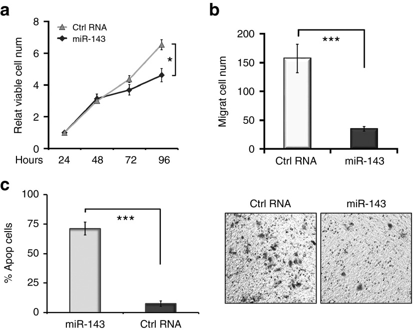
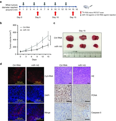
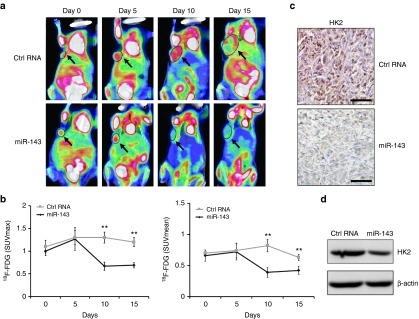
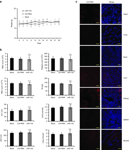
Increased glucose utilization is a hallmark of cancer, and tumor metabolism is emerging as anticancer target for therapeutic intervention. Triple-negative breast cancers TNBC are highly glycolytic and show poor clinical outcomes. We previously identified hexokinase 2, the major glycolytic enzyme, as a target gene of miR-143 in TNBC. Here, we developed a therapeutic formulation using cholesterol-modified miR-143 agomir encapsulated in a neutral lipid-based delivery agent that blocked tumor growth and glucose metabolism in TNBC tumor-bearing mice when administered systemically. The antioncogenic effects were accompanied by a reduction in the direct target hexokinase 2 and [(18)F]-fluorodeoxyglucose ((18)F-FDG) uptake based on positron emission tomography/computed tomography. Treatment with miR-143 formulation has minimal toxic effects and mice tolerated it well. Thus, we demonstrated that miR-143 is a robust inhibitor of the Warburg effect and an effective therapeutic target for TNBC. In addition, (18)F-FDG positron emission tomography/computed tomography can be used to specifically monitor the response of TNBC to miR-143-based therapeutics by targeting tumor glycolysis.


Figures

Figure 1
Figure 1
miR-143 represses glycolysis by targeting hexokinase 2 (HK2) in MDA-MB-231 cells. (a) Following transfection with RNA oligonucleotides (miR-143 mimic or Ctrl RNA) and incubation in low-glucose medium, the glucose metabolism rates of MDA-MB-231 cells were detected using specialized kits. The left panel shows the rates of glucose consumption, while the right panel shows the rates of lactate production. (b) miR-143 repressed [18F]-fluorodeoxyglucose (18F-FDG) uptake of MDA-MB-231 cells in an in vitro dynamic 18F-FDG uptake assay. (c) miR-143 downregulated the mRNA level of HK2 in quantitative reverse-transcriptase-polymerase chain reaction (qRT-PCR). Mock represents the phosphate buffered saline (PBS) group. (d) miR-143 inhibited protein expression of HK2 as evident by western blotting. Values represent the mean ± SD of three separate experiments. *P < 0.05, **P < 0.01, ***P < 0.001.
Figure 2
Figure 2
miR-143 regulates the proliferation, migration, and apoptosis of MDA-MB-231 cells. (a) miR-143 inhibited the proliferation activity of MDA-MB-231 cells in MTT assays. (b) miR-143 effectively suppressed cell migration in Transwell migration assays. (c) miR-143 significantly induced cell apoptosis in flow cytometry assays. Values represent the mean ± SD of three separate experiments. *P < 0.05, **P < 0.01, ***P < 0.001.
Figure 3
Figure 3
Systemic delivery of miR-143 agomir inhibits tumor growth in mouse models of triple-negative breast cancer (TNBC). (a) Schematic diagram of the experimental design. For this experiment, 6–8-week-old female BALB/c athymic nude mice were used for subcutaneous xenografts. When tumor diameters reached ~ 5 mm, Cy3-labeled miR-143 agomir (treatment group) or Cy3-labeled Ctrl RNA agomir (negative control group) encapsulated in a lipid-based delivery vehicle was administered through tail vein injections at 1.5 mg/kg of body weight every 3 days for 5 cycles. (b) Time course of TNBC tumor growth in mice treated with miR-143 agomir or Ctrl RNA agomir. (c) Tumors were resected from miR-143 agomir and Ctrl RNA agomir mice. (d) Cy3 signals were detectable in tumor tissue slices from Cy3-labeled miRNA agomir shown in the upper left panels. Nucleus counterstained with 4′,6-diamidino-2-phenylindole on tumor tissue slices are shown in the middle left panels. The merged images are shown in the lower left panels. Immunohistochemical staining is presented in the right panels and included hematoxylin and eosin (HE), proliferating cell nuclear antigen (PCNA) (brown), and caspase-3 (brown) detection of TNBC tumor sections from the 2 groups. Scale bars: 100 μm. All data are mean ± SD of three separate experiments. *P < 0.05, **P < 0.01, ***P < 0.001. n = 3 in each group.
Figure 4
Figure 4
Assessment of response to miR-143-based therapy in triple-negative breast cancer (TNBC) xenografts. (a) [18F]-fluorodeoxyglucose (18F-FDG) microPET/CT imaging of mice treated with miR-143 agomir or Ctrl RNA agomir at days 0 (baseline), 5, 10, and 15. Representative 18F-FDG micro positron emission tomography/computed tomography (PET/CT) images are shown with circles indicating xenografted TNBC tumors. (b) Quantification of tumor uptake of 18F-FDG is presented as maximum standardized uptake value (SUVmax) and mean standardized uptake value (SUVmean). (c) Hexokinase2 (HK2) expression of TNBC tumor sections from immunohistochemical staining. (d) HK2 protein levels determined by western blotting. All data are mean ± SD of three separate experiments. *P < 0.05, **P < 0.01, ***P < 0.001.
Figure 5
Figure 5
Toxicity analyses following intravenous delivery of encapsulated miRNA agomir in triple-negative breast cancer (TNBC) xenografts. Another three randomized groups of TNBC tumor-bearing xenografts were established (n = 3 mice in each group). When tumor diameters reached ~5 mm, encapsulated miRNA agomirs were administered by tail vein injection at 1.5 mg/kg of body weight every 3 days for 10 cycles. (a) Body weights were measured every 3 days throughout the study. (b) Routine blood tests and biochemical tests were performed in normal BALB/c athymic nude mice after treatment with 1.5 mg/kg of body weight miR-143 agomir or Ctrl RNA agomir or phosphate buffered saline (PBS). (c) Cy3 signals were examined in heart, lung, brain, liver, kidney, spleen, muscle tissue slices shown in the left panels to evaluate the biodistribution of Cy3-labeled miR-143 agomir. The merged images with 4′,6-diamidino-2-phenylindole nucleus counterstained are shown in the right panels. Scale bars: 50 μm. Data are presented as mean ± SD.

References

    1. Torre, LA, Bray, F, Siegel, RL, Ferlay, J, Lortet-Tieulent, J and Jemal, A (2015). Global cancer statistics, 2012. CA Cancer J Clin 65: 87–108. - PubMed
    1. Carey, L, Winer, E, Viale, G, Cameron, D and Gianni, L (2010). Triple-negative breast cancer: disease entity or title of convenience? Nat Rev Clin Oncol 7: 683–692. - PubMed
    1. Kassam, F, Enright, K, Dent, R, Dranitsaris, G, Myers, J, Flynn, C et al. (2009). Survival outcomes for patients with metastatic triple-negative breast cancer: implications for clinical practice and trial design. Clin Breast Cancer 9: 29–33. - PubMed
    1. Lehmann, BD, Bauer, JA, Chen, X, Sanders, ME, Chakravarthy, AB, Shyr, Y et al. (2011). Identification of human triple-negative breast cancer subtypes and preclinical models for selection of targeted therapies. J Clin Invest 121: 2750–2767. - PMC - PubMed
    1. Hanahan, D and Weinberg, RA (2011). Hallmarks of cancer: the next generation. Cell 144: 646–674. - PubMed

LinkOut - more resources